. 2022 Dec 24;15(1):58.
doi: 10.3390/v15010058.

The Interaction of Mandarin Fish DDX41 with STING Evokes type I Interferon Responses Inhibiting Ranavirus Replication

Affiliations

The Interaction of Mandarin Fish DDX41 with STING Evokes type I Interferon Responses Inhibiting Ranavirus Replication

Xiao-Wei Qin et al. Viruses. .

Abstract

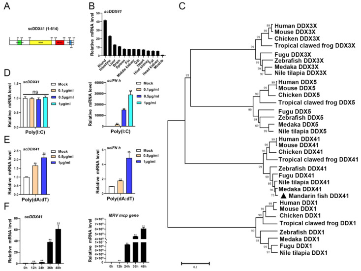
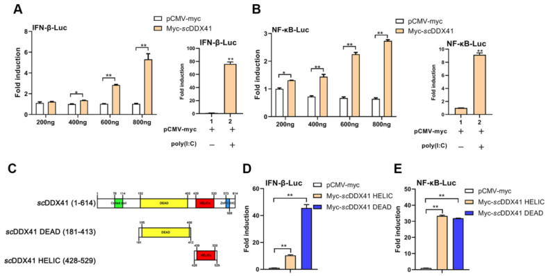
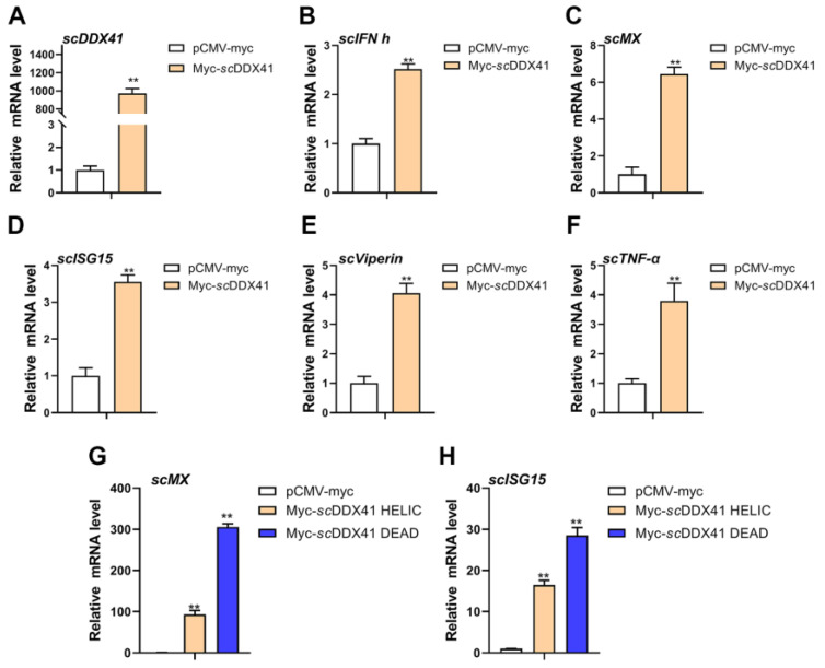
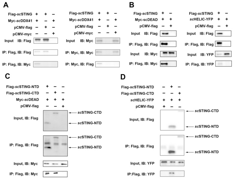
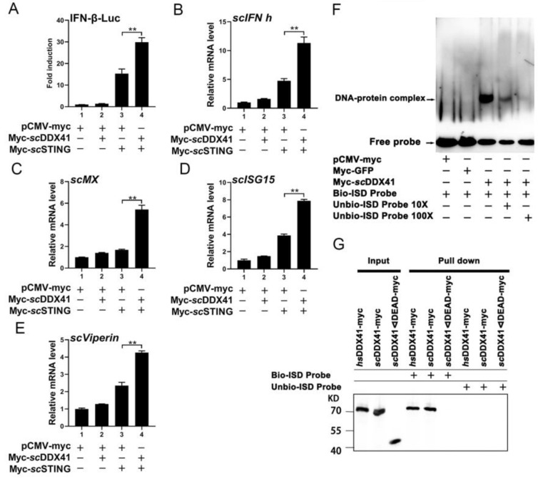
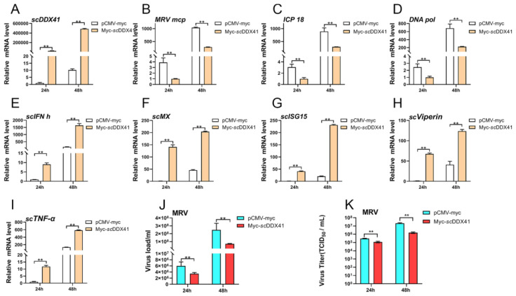
DDX41 is an intracellular DNA sensor that evokes type I interferon (IFN-I) production via the adaptor stimulator of interferon gene (STING), triggering innate immune responses against viral infection. However, the regulatory mechanism of the DDX41-STING pathway in teleost fish remains unclear. The mandarin fish (Siniperca chuatsi) is a cultured freshwater fish species that is popular in China because of its high market value. With the development of a high-density cultural mode in mandarin fish, viral diseases have increased and seriously restricted the development of aquaculture, such as ranavirus and rhabdovirus. Herein, the role of mandarin fish DDX41 (scDDX41) and its DEAD and HELIC domains in the antiviral innate immune response were investigated. The level of scDDX41 expression was up-regulated following treatment with poly(dA:dT) or Mandarin fish ranavirus (MRV), suggesting that scDDX41 might be involved in fish innate immunity. The overexpression of scDDX41 significantly increased the expression levels of IFN-I, ISGs, and pro-inflammatory cytokine genes. Co-immunoprecipitation and pull-down assays showed that the DEAD domain of scDDX41 recognized the IFN stimulatory DNA and interacted with STING to activate IFN-I signaling pathway. Interestingly, the HELIC domain of scDDX41 could directly interact with the N-terminal of STING to induce the expression levels of IFN-I and ISGs genes. Furthermore, the scDDX41 could enhance the scSTING-induced IFN-I immune response and significantly inhibit MRV replication. Our work would be beneficial to understand the roles of teleost fish DDX41 in the antiviral innate immune response.

Keywords: DDX41; MRV; STING; innate immune; interferon; mandarin fish; ranavirus.


Conflict of interest statement

The authors declare no conflict of interest.

Figures

Figure 1
Figure 1
(A) SMART analysis of DDX41s. Coiled coil domain, DEAD domain, HELIC domain, and ZnF_C2HC domain were labeled in the sequence. (B) Distributions of scDDX41 expression in different tissues of mandarin fish. The expression levels of scDDX41 were detected by RT-qPCR. (C) Phylogenetic tree of DDX41 proteins from various species. A phylogenetic tree was constructed using the Neighbor-Joining method in MEGA v10.0, with 1000 bootstrap replications. The bootstrap values were indicated at the nodes of the tree. (D) Expression levels of scDDX41 in cells treated with poly(I:C) at indicated times. (E) Expression levels of scDDX41 in cells treated with poly (dA:dT) at indicated times. (F) Expression levels of scDDX41 in cells infected with MRV at indicated times. The β-actin gene served as internal control to calibrate the cDNA template for all samples. Vertical bars represent ±SD (n = 3). Statistical significance was indicated by asterisks, with ** referring to p < 0.01. ns, non-significant.
Figure 2
Figure 2
scDDX41 induced the activities of IFN-β-luc and NF-κB-luc promoters. (A) Cells were transfected with 0.2, 0.4, 0.6, or 0.8 μg of scDDX41 expression plasmid or an empty vector together with IFN-β-luc (0.4 μg/well) and pRL-TK (0.04 μg/well) plasmids. Luciferase assays were performed 36 h after the transfection. (B) Cells were transfected with 0.2, 0.4, 0.6, or 0.8 μg of scDDX41 expression plasmid or an empty vector together with NF-κB-luc (0.4 μg/well) and pRL-TK (0.05 μg/well). Luciferase assays were performed 36 h after the transfection. (C) Schematic of full-length and scDDX41 mutants with the DEAD domain, HELIC domain, and residue numbers as indicated. Various scDDX41 fragments were inserted into the C-terminus of pCMV-myc. (D,E) DEAD and HELIC domains of scDDX41 for IFN and NF-κB activation. Cells were transfected with 0.4 μg/well of various expression plasmids of scDDX41, scDDX41 mutants, or empty vector together with the reporter plasmid 0.04 μg/well pRL-TK as well as 0.4 μg/well of IFN-β-luc or NF-κB-luc plasmid. Luciferase assays were performed 36 h after the transfection. All luciferase assays were repeated at least three times, and data are means ±SD (n = 3) from single representative experiments. * p < 0.05, ** p < 0.01 between normal cells and stimulated cells.
Figure 3
Figure 3
Overexpression of scDDX41 induces the expression of IFN-I, ISGs, and inflammatory cytokines. After transfection with scDDX41-myc or pCMV-myc at 24 h, cells were harvested and the expression levels of scDDX41 (A), scIFN-h (B), scMx (C), scISG15 (D), scViperin (E) and scTNF-α (F) genes were detected. Overexpression of scHELIC and scDEAD induced the expression of scMx (G) and scISG15 (H) in MFF-1 cells. The β-actin gene served as the internal control to calibrate the cDNA template for all samples. Vertical bars represent ±SD (n = 3). Statistical significance is indicated by asterisks, with ** referring to p < 0.01.
Figure 4
Figure 4
Identification of the interaction between scDDX41 and scSTING by Co-IP assay. (A) scDDX41 interacted with scSTING. Cells were transfected with the indicated plasmids. At 36 h post-transfection, the cell lysates were precipitated with an anti-flag or anti-myc mAb in conjunction with protein G-Sepharose beads and detected by WB analysis using anti-myc or anti-flag mAbs. The expression of the transfected proteins was analyzed by immunoblotting with anti-myc and anti-flag mAbs. (B) DEAD and HELIC domains of scDDX41 interacted with scSTING. Cells were co-transfected with DEAD-myc, HELIC-YFP, and scSTING-flag or empty vector. Immunoprecipitation assays with anti-flag antibody (IP: Flag) and Western blot analysis were performed with anti-Flag, anti-myc, or anti-YFP antibodies. (C) DEAD domain interacted with scSTING-NTD and scSTING-CTD. Cells were co-transfected with myc-DEAD and flag-scSTING-NTD, flag-scSTING-CTD, or empty vector. Immunoprecipitation assays with anti-flag antibody (IP: Flag) and WB analysis were performed with anti-Flag or anti-myc antibodies. (D) HELIC domain interacted with scSTING-NTD but not with scSTING-CTD. Cells were co-transfected with HELIC-YFP and flag-scSTING-NTD, flag-scSTING-CTD, or empty vector. Immunoprecipitation assays with anti-flag antibody (IP: Flag) and WB analysis were performed with anti-flag or anti-YFP antibodies.
Figure 5
Figure 5
Involvement of scDDX41 in scSTING-mediated IFN expression, and scDDX41 recognizes dsDNA through the DEAD domain. (A) Cells transfected with an IFN-β-luc 400 ng/well and TK (40 ng/well) plus 400 ng/well) of the expression vectors for pCMV-myc, pCMV-myc and scDDX41-myc, pCMV-myc and scSTING-myc, scDDX41-myc, and scSTING-myc. Vertical bars represent ±SD (n = 3). Statistical significance is indicated by asterisks, with ** referring to p < 0.01. scDDX41 enhanced the IFN-I response induced by STING. (BE) Cells seeded in 6-well plates were transfected or co-transfected with scDDX41 (400 ng/well), and scSTING (400 ng/well). The cells transfected with pCMV-myc acted as negative control. The expression levels of interferon signaling molecules including scIFN-h, scMx, scISG15, and scViperin were examined using RT-qPCR. The β-actin gene served as the internal control to calibrate the cDNA template for all samples. Vertical bars represent ±SD (n = 3). Statistical significance is indicated by asterisks, with ** referring to p < 0.01. (F) scDDX41 recognizes ISD through the DEAD domain using EMSA with biotin-labeled (Bio-) or unlabeled (Unbio-) probes ISD. The black lines indicate where parts of the image were joined. (G) Immunoblot analysis of the immunoprecipitated purified myc-tagged hsDDX41, scDDX41, or scDDX41ΔDEAD recombinant proteins incubated individually with biotinylated ISD and probed with anti-myc antibodies.
Figure 6
Figure 6
Overexpression of scDDX41 attenuates MRV infection. After transfection with scDDX41-myc or pCMV-myc at 24 h, cells were infected with MRV and harvested for RT-qPCR independently at the indicated time points. (A) Expression levels of scDDX41 in cells infected with MRV at indicated times. (BD) Expression levels of mcp, ICP-18, and DNA pol genes in cells infected with MRV at indicated times. (EI) Expression levels of scIFN-h, scMx, scISG15, scViperin, and scTNF-α genes in cells infected with MRV at indicated times. (J) Correlation of viral load in samples measured by TaqMan qPCR. (K) The titer of virus infection was measured on a 96-well cell culture plate via the finite dilution method. β-actin gene served as the internal control to calibrate the cDNA template for all samples. Vertical bars represent ±SD (n = 3). Statistical significance is indicated by asterisks, with ** referring to p < 0.01.

Similar articles

Cited by

References

    1. Linder P., Fullerpace F.V. Looking back on the birth of DEAD-box RNA helicases. Biochim. Biophys. Acta. 2013;1829:750–755. doi: 10.1016/j.bbagrm.2013.03.007. - DOI - PubMed
    1. Kadono M., Kanai A., Nagamachi A., Shinriki S., Kawata J., Iwato K., Kyo T., Oshima K., Yokoyama A., Kawamura T. Biological implications of somatic DDX41 p.R525H mutation in acute myeloid leukemia. Exp. Hematol. 2016;44:745–754. doi: 10.1016/j.exphem.2016.04.017. - DOI - PubMed
    1. Omura H., Oikawa D., Nakane T., Kato M., Ishii R., Ishitani R., Tokunaga F., Nureki O. Structural and Functional Analysis of DDX41: A bispecific immune receptor for DNA and cyclic dinucleotide. Sci. Rep. 2016;6:34756. doi: 10.1038/srep34756. - DOI - PMC - PubMed
    1. Schmucker D., Jackle H., Gaul U. Genetic analysis of the larval optic nerve projection in Drosophila. Development. 1997;124:937–948. doi: 10.1242/dev.124.5.937. - DOI - PubMed
    1. Lee K.G., Kim S.S.Y., Kui L., Voon D.C.C., Mauduit M., Bist P., Bi X., Pereira N.A., Liu C., Sukumaran B. Bruton’s Tyrosine Kinase Phosphorylates DDX41 and Activates Its Binding of dsDNA and STING to Initiate Type 1 Interferon Response. Cell Rep. 2015;10:1055–1065. doi: 10.1016/j.celrep.2015.01.039. - DOI - PubMed

Publication types