. 2022 Dec 30;15(1):114.
doi: 10.3390/v15010114.

Mouse Adapted SARS-CoV-2 (MA10) Viral Infection Induces Neuroinflammation in Standard Laboratory Mice

Affiliations

Mouse Adapted SARS-CoV-2 (MA10) Viral Infection Induces Neuroinflammation in Standard Laboratory Mice

Narayanappa Amruta et al. Viruses. .

Abstract

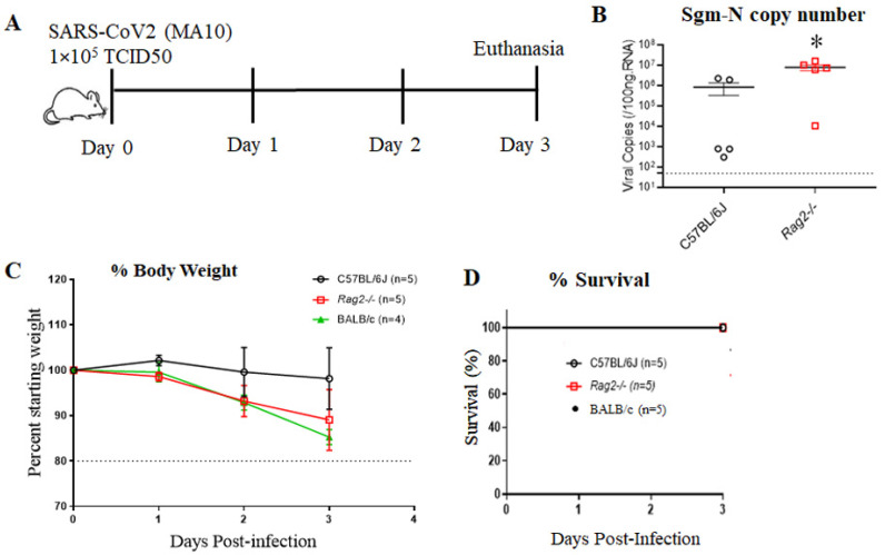
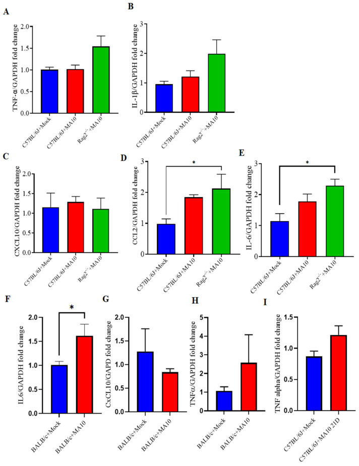
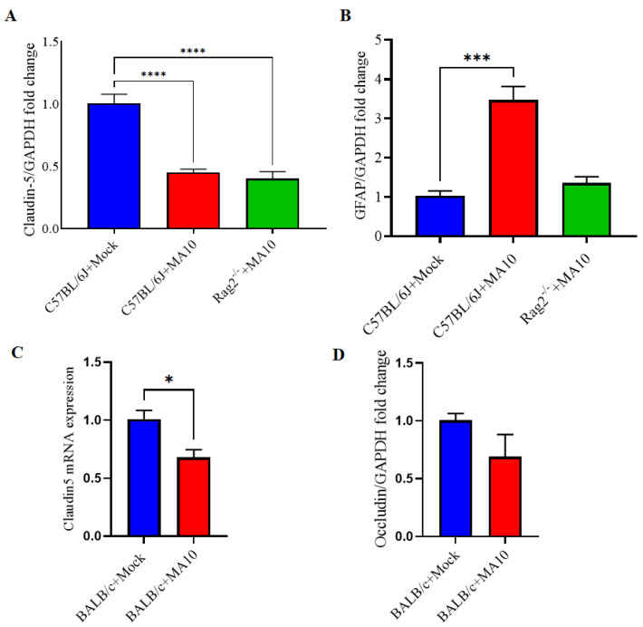
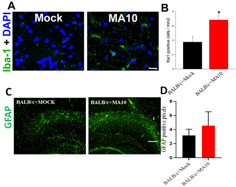
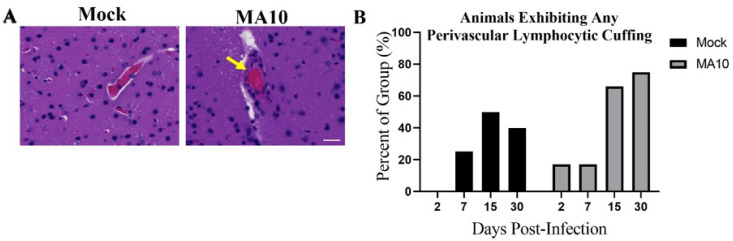
Increasing evidence suggests that severe acute respiratory syndrome coronavirus 2 (SARS-CoV-2) infection impacts neurological function both acutely and chronically, even in the absence of pronounced respiratory distress. Developing clinically relevant laboratory mouse models of the neuropathogenesis of SARS-CoV-2 infection is an important step toward elucidating the underlying mechanisms of SARS-CoV-2-induced neurological dysfunction. Although various transgenic models and viral delivery methods have been used to study the infection potential of SARS-CoV-2 in mice, the use of commonly available laboratory mice would facilitate the study of SARS-CoV-2 neuropathology. Herein we show neuroinflammatory profiles of immunologically intact mice, C57BL/6J and BALB/c, as well as immunodeficient (Rag2-/-) mice, to a mouse-adapted strain of severe acute respiratory syndrome coronavirus-2 (SARS-CoV-2 (MA10)). Our findings indicate that brain IL-6 levels are significantly higher in BALB/c male mice infected with SARS-CoV-2 MA10. Additionally, blood-brain barrier integrity, as measured by the vascular tight junction protein claudin-5, was reduced by SARS-CoV-2 MA10 infection in all three strains. Brain glial fibrillary acidic protein (GFAP) mRNA was also elevated in male C57BL/6J infected mice compared with the mock group. Lastly, immune-vascular effects of SARS-CoV-2 (MA10), as measured by H&E scores, demonstrate an increase in perivascular lymphocyte cuffing (PLC) at 30 days post-infection among infected female BALB/c mice with a significant increase in PLC over time only in SARS-CoV-2 MA10) infected mice. Our study is the first to demonstrate that SARS-CoV-2 (MA10) infection induces neuroinflammation in laboratory mice and could be used as a novel model to study SARS-CoV-2-mediated cerebrovascular pathology.

Keywords: COVID-19; SARS-CoV-2; animal models; brain; mouse-adaptation; neuroinflammation.


Conflict of interest statement

The authors declare no conflict of interest.

Figures

Figure 1
Figure 1
C57BL/6J mice show less disease severity compared to BALB/c and Rag2−/− mice following mouse-adapted SARS-CoV2 (MA10) infection. (A) Schematic overview of the experimental timeline for 10-week-old male C57BL/6, BALB/c, and Rag2−/− mice. Animals were inoculated via the intranasal route with mock (PBS) or MA10 strain of SARS-CoV-2 (1 × 10⁵ TCID50). Upon 3 days post-infection (3 dpi), mice were euthanized, and RNA isolated from the lung samples by the Trizol method was processed for gene expression. (B) Subgenomic-N viral copies and (C) Percent starting weight loss in SARS-CoV-2 (MA10)-infected C57BL/6 and SARS-CoV-2 (MA10) infected Rag2−/− mice and BALB/c (D) Survival rate. Data were analyzed using 2-factor ANOVA followed by Sidak’s multiple comparisons if more than two groups. The Mann-Whitney U test is used to compare the difference in N viral copies between C57Bl/6J and Rag2−/−. Error bars represent mean +/− SEM, * p < 0.05, n = 4–5/group.
Figure 2
Figure 2
CNS cytokine and chemokine responses in the brains of mouse-adapted SARS-CoV-2 (MA10) infected laboratory mice. 10-week-old male C57BL/6, BALB/c and Rag2−/− mice were inoculated via the intranasal route with mock or MA10 strain of SARS-CoV-2 (1 × 10⁵ TCID50). Upon 3 days post-infection (3 dpi), mice were euthanized, and RNA was isolated from the left hemisphere for gene expression analysis. (A) TNF-α and (B) IL-1β mRNA expression in brains of SARS-CoV-2 (MA10) infected C57BL/6J mice did not significantly differ from those of mock-infected mice. (C) Cxcl10; (D) Ccl2 and (E) IL-6 expression in brains of SARS-CoV-2 (MA10) infected Rag2−/− mice and C57BL/6J mock-treated mice. (F) IL-6 (G) CxCL10 (H) TNF-α expression in BALB/c mice. (I) 21 days post-infection (21 dpi), male mice were euthanized and analyzed for TNF-α mRNA expression in C57BL/6 mice, SARS-CoV-2 MA10 infected C57BL/6 mice show a trend towards increased TNF-α expression compared to mock-treated mice. IL-6 was significantly increased, and TNF-α showed an upward trend in SARS-CoV-2 MA10-infected BALB/c mice compared with mock-treated mice. Data are presented as mean ± SEM. p values represent mock vs. SARS-CoV-2 (MA10) challenged groups. Significant differences are designated using a two-tailed unpaired student t-test for two groups and one-way ANOVA (for more than two groups). Some of the experiment n = 4 indicates 2 mice were excluded because the dissection of half left hemisphere did not align with other mice. C57BL/6J + Mock n = 5; C57BL/6J + SARS-CoV-2 MA10 n = 4; BALB/c + Mock n = 4–5; BALB/c + SARS-CoV-2 MA10 n = 4–5; Rag2−/− + SARS-CoV-2 MA10 n = 4; * p < 0.05.
Figure 3
Figure 3
mRNA expression of Claudin-5 is decreased, and GFAP increased in the brains of mouse-adapted SARS-CoV-2 (MA10) infected mice. 10-week-old male C57BL/6J, BALB/c and Rag2−/− mice were inoculated via the intranasal route with mock or MA10 strain of SARS-CoV-2 (1 × 10⁵ TCID50; Red bar). Upon 3 days post-infection (3 dpi), mice were euthanized, and RNA was isolated from the half-left hemisphere of the brain by the Trizol method for gene expression analysis. (A) Claudin-5 and (B) GFAP mRNA expression in brains of mock or SARS-CoV-2 (MA10) infected C57BL/6 mice, whereas (C) Claudin-5 (D) Occludin expression in BALB/c mice were evaluated. Claudin-5 expression was significantly lower in brains of all SARS-CoV-2 (MA10) infected C57BL/6J, BALB/c and Rag2−/− mice compared with C57BL/6J and BALB/c mock-treated mice. GFAP expression was significantly higher in the brains of SARS-CoV-2 (MA10) infected C57BL/6J mice. Occludin expression (D) showed a downtrend in SARS-CoV-2 MA10-infected BALB/c mice compared with mock-treated mice. Data are presented as mean ± SEM. p values represent mock vs. SARS-CoV-2 (MA10) challenged groups. Significant differences are designated using a two-tailed unpaired student t-test for two groups and one-way ANOVA (for more than two groups). C57BL/6J + Mock n = 5; C57BL/6J + SARS-CoV-2 MA10 n = 5; Rag2−/− + SARS-CoV-2 MA10 n = 5; * p < 0.05, *** p < 0.001, **** p < 0.0001.
Figure 4
Figure 4
SARS-CoV-2 (MA10) infection significantly increases Iba-1 positive microglial cells in the cortex region of the brain in 1-year-old female BALB/c mice. 1-year-old female BALB/c mice were inoculated via the intranasal route with saline (black bar) or SARS-CoV-2 (MA10) strain of SARS-CoV-2 (1 × 10³ PFU/mL) (Red bar) (Detailed experimental methods are available in Dinnon et al., 2020; Leist et al., 2021). Upon 2 days post-infection (2 dpi), mice were euthanized, and the whole brain was harvested and fixed in 10% phosphate-buffered formalin, paraffin-embedded and sectioned at 4μm thickness. Sequential sections were stained with Iba-1 by immunofluorescence. (A) Representative images of Iba-1 positive microglial cells (Iba1 staining, green) with DAPI as nuclear counterstaining show high induction in the cortex region of brains of SARS-CoV-2 (MA10) infected female BALB/c mice. (B) Quantification of Iba-1 positive microglial cells was significantly higher in SARS-CoV-2 (MA10) infected brains compared with mock infection. SARS-CoV-2 (MA10) infection increases (C,D) GFAP-positive cells in the hippocampus of 12-week old male BALB/c mice after 3 days of infection. Data are presented as mean ± SEM. p values represent saline vs SARS-CoV-2 (MA10) challenged groups. Significant differences are designated using a two-tailed unpaired student t-test. Saline n = 5; SARS-CoV-2 MA10 n = 6, * p < 0.01. Scale bar: 40× magnification, scale bar  =  25 μm.
Figure 5
Figure 5
SARS-CoV-2 (MA10) infection increases perivascular lymphocyte cuffing in the brains of BALB/c mice. 1-year-old female mice were infected as in Figure 3. (A) Representative H&E-stained brain images as labeled demonstrate significant perivascular lymphocyte cuffing (yellow arrowheads) at 30 days (30 dpi) post-infection SARS-CoV-2 (MA10) infected female mice. Scale bar = 20 μm. (B) Quantification of the percent of each group of animals (Saline n = 4; MA10 n = 6) with any detectable perivascular lymphocyte cuffing at various days post-infection as labeled. A total of saline n = 16 and MA10 n = 24 were included in the study. Saline n = 4; MA10 n = 6 was euthanized at each time point (each group of animals), and all mice were undergone for histological analysis. Graphs represent only the percent of each group of animals that showed positive for histopathological symptoms of perivascular lymphocyte cuffing. Chi-Square trend analysis shows a significant change in the portion of mice with detectable perivascular lymphocytic cuffing over time only in SARS-CoV-2 (MA10)-infected mice.
Figure 6
Figure 6
Intranasal administration of SARS-CoV-2 (MA10) upon accumulation in the mice upregulates Ccl2 and Il-1β, activates microglia, induces perivascular lymphocyte cuffing, promotes astrocyte accumulation, triggers a loss of tight junction protein claudin-5. These neuroinflammatory events mediate cerebrovascular pathology in the mice infected with SARS-CoV-2 (MA10). (https://biorender.com/ accessed on 30 October 2022).

References

    1. Amruta N., Chastain W.H., Paz M., Solch R.J., Murray-Brown I.C., Befeler J.B., Gressett T.E., Longo M.T., Engler-Chiurazzi E.B., Bix G. SARS-CoV-2 mediated neuroinflammation and the impact of COVID-19 in neurological disorders. Cytokine Growth Factor Rev. 2021;58:1–15. doi: 10.1016/j.cytogfr.2021.02.002. - DOI - PMC - PubMed
    1. Sanyaolu A., Okorie C., Marinkovic A., Patidar R., Younis K., Desai P., Hosein Z., Padda I., Mangat J., Altaf M. Comorbidity and its Impact on Patients with COVID-19. SN Compr. Clin. Med. 2020;2:1069–1076. doi: 10.1007/s42399-020-00363-4. - DOI - PMC - PubMed
    1. Klein R., Soung A., Sissoko C., Nordvig A., Canoll P., Mariani M., Jiang X., Bricker T., Goldman J., Rosoklija G., et al. COVID-19 induces neuroinflammation and loss of hippocampal neurogenesis. Res. Sq. 2021 doi: 10.21203/rs.3.rs-1031824/v1. - DOI - PubMed
    1. Wang Q., Davis P.B., Gurney M.E., Xu R. COVID-19 and dementia: Analyses of risk, disparity, and outcomes from electronic health records in the US. Alzheimers Dement. 2021;17:1297–1306. doi: 10.1002/alz.12296. - DOI - PMC - PubMed
    1. Wu Y., Xu X., Chen Z., Duan J., Hashimoto K., Yang L., Liu C., Yang C. Nervous system involvement after infection with COVID-19 and other coronaviruses. Brain Behav. Immun. 2020;87:18–22. doi: 10.1016/j.bbi.2020.03.031. - DOI - PMC - PubMed

Publication types