. 2022 Dec 31;15(1):139.
doi: 10.3390/v15010139.

Monoclonal Antibodies Specific for SARS-CoV-2 Spike Protein Suitable for Multiple Applications for Current Variants of Concern

Affiliations

Monoclonal Antibodies Specific for SARS-CoV-2 Spike Protein Suitable for Multiple Applications for Current Variants of Concern

Mahali S Morgan et al. Viruses. .

Abstract

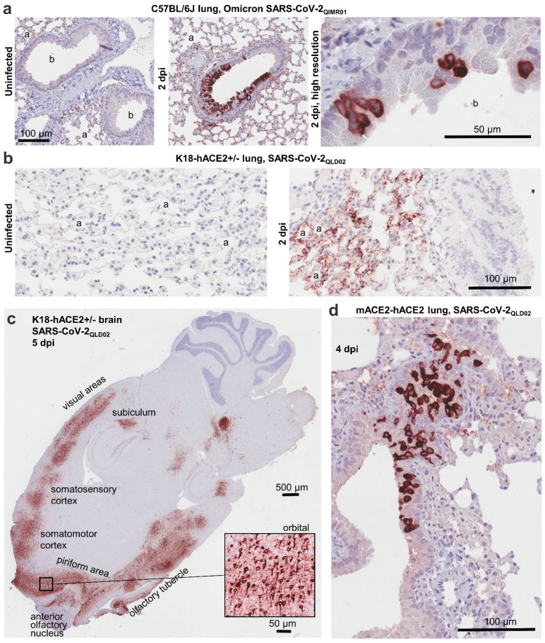
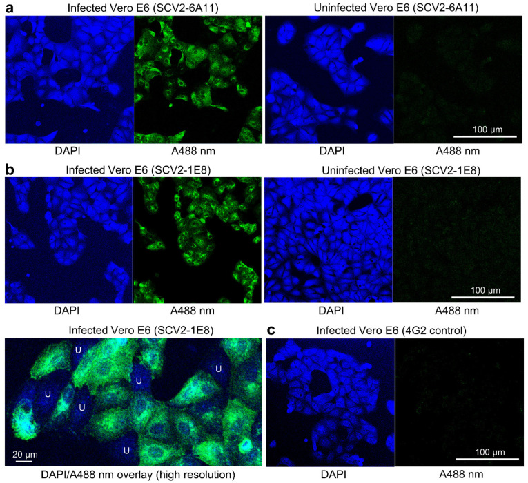
The global coronavirus disease 2019 (COVID-19) pandemic caused by the severe acute respiratory syndrome coronavirus 2 (SARS-CoV-2) has spawned an ongoing demand for new research reagents and interventions. Herein we describe a panel of monoclonal antibodies raised against SARS-CoV-2. One antibody showed excellent utility for immunohistochemistry, clearly staining infected cells in formalin-fixed and paraffin embedded lungs and brains of mice infected with the original and the omicron variants of SARS-CoV-2. We demonstrate the reactivity to multiple variants of concern using ELISAs and describe the use of the antibodies in indirect immunofluorescence assays, Western blots, and rapid antigen tests. Finally, we illustrate the ability of two antibodies to reduce significantly viral tissue titers in K18-hACE2 transgenic mice infected with the original and an omicron isolate of SARS-CoV-2.

Keywords: SARS-CoV-2; Western blotting; immunofluorescence; immunohistochemistry; immunotherapy; lateral flow assays; monoclonal antibody.


Conflict of interest statement

One or more of the mAbs may be sold under commercial agreement(s) with some revenue returning to the authors.

Figures

Figure 1
Figure 1
Immunohistochemistry using SCV2-1E8. (a) Uninfected lungs (left) and infected lungs at 2 dpi (two images on the right) from C57BL/6J mice infected with an omicron isolate, SARS-CoV-2QIMR01. (b) Uninfected lungs (left) and infected lungs at 2 dpi (right) from K18-hACE2+/− mice infected with an original strain isolate, SARS-CoV-2QLD02. (c) Brain from K18-hACE2+/− mice on 5 dpi with SARS-CoV-2QLD02. Stained areas of the brain are annotated (approximate); insert taken from the orbital region of the brain. (d) Infected lungs 4 dpi from mACE2-hACE2 mice infected with SARS-CoV-2QLD02. Within the IHC panels of (a) and (b), alveolar spaces (a) and bronchioles (b) are indicated.
Figure 2
Figure 2
Indirect Immunofluorescence Assay (IFA) staining. (a) Vero E6 cells infected with SARS-CoV-2QLD02 (left) or uninfected (right) stained with DAPI (blue) and anti-SARS-CoV-2 mAb, SCV2-6A11 (green, A488 nm). (b) As in a, but stained with SCV2-1E8. Bottom left shows a high resolution image with DAPI/A488 nm overlap, where infected cells (green) can clearly be distinguished from uninfected cells (blue, labelled U). (c) Vero E6 cells infected with SARS-CoV-2QLD02 stained with a negative control mAb, 4G2.
Figure 3
Figure 3
ELISAs and Western blots. (a) ELISA curves for the indicated purified mAbs using recombinant HexaPro spike proteins with mutated furin cleavage site from the indicated variants of concern as antigen. (b) Western blots using reduced (DDT) lysates of Vero E6 cells with (+) and without (−) infection with SARS-CoV-2QLD02 (left), or lysates of SARS-CoV-2QLD02 infected Vero E6 cells that were either reduced (+) or not reduced (−) (right). The primary reactivity of each mAb is indicated. (Note SCV2-6A11 is used in both settings).
Figure 4
Figure 4
Lateral flow spike antigen detection applications. (a) Setup of the lateral flow spike antigen detection system (generated using BioRender). Sample (containing recombinant spike) is applied followed by running buffer, which carries the antigen and mobilizes the gold-conjugated SCV2-7E9 mAb. Spike bound to SCV-6A11 (the capture mAb) is then detected by SCV2-7E9-Gold. The control binds the SCV2-7E9-Gold illustrating that the detection-mAb has been mobilized. (b) The SCV2-7E9-Gold is visualized as a red band that illustrates the presence of recombinant spike in the sample (TEST), with a red CONTROL line illustrating that the detection mAb (SCV2-7E9-Gold) has been mobilized. (c) Quantitation of antigen detection by the lateral flow test for recombinant spike antigens from each of 3 variants of concern. (SD from n = 3 replicates).
Figure 5
Figure 5
Virus neutralization assays. The indicated concentrations of purified antibodies were serially diluted in duplicate and used to neutralize SARS-CoV-2QLD02 in Vero E6 CPE-based assays. High OD values illustrate cells protected from CPE by mAbs and thus staining with crystal violet, and low OD values represents a loss of cells via CPE resulting in loss of crystal violet staining; 50% inhibitory concentrations (IC50) were determined by interpolation. 100% CPE (0% neutralization) was determined in 8 replicates in wells without mAb. IC50 values were determined using the “log(inhibitor) vs. response” feature of GraphPad Prism (purple).
Figure 6
Figure 6
Protection in K18-ACE2 mice. (a) Timeline of mAb therapy of K18-ACE2+/+ mice infected with a BA.1 omicron isolate (SARS-CoV-2QIMR01). The anti-SARS-CoV-2 antibody was SCV2-5A1 and the control mAb was an isotype control, anti-Zika virus NS1 (3H3). (b) Lung and nasal turbinate titers for mice described in a. Statistics by t test (lung) and Kolmogorov–Smirnov test (nasal turbinates). (c) Timelines for prophylactic and therapeutic treatment of K18-ACE2+/− mice infected with an original strain isolate (SARS-CoV-2QLD02) with SCV2-5A1 or SCV2-3H9, or the control mAb, anti-flavivirus E (4G2). (d) Mean weight change relative to 0 dpi after mAb therapy. Statistics by Kolmogorov–Smirnov tests for 5 dpi. (e) Tissue titrations for the same mice shown in d (5 dpi). Statistics by Kolmogorov–Smirnov tests. N.S.—not significant. (f) Mean weight change relative to 0 dpi after mAb prophylaxis. For statistics, data for 4G2 and PBS were combined to represent the controls (n = 5). Statistics by Kolmogorov–Smirnov tests on 5 dpi. (g) Tissue titrations for the same mice are shown in f (5 dpi). Statistics by Kolmogorov–Smirnov tests, with 4G2 and PBS groups combined.

References

    1. McHenry A., Iyer K., Wang J., Liu C., Harigopal M. Detection of SARS-CoV-2 in tissue: The comparative roles of RT-qPCR, in situ RNA hybridization, and immunohistochemistry. Expert Rev. Mol. Diagn. 2022;22:559–574. doi: 10.1080/14737159.2022.2085508. - DOI - PubMed
    1. Best Rocha A., Stroberg E., Barton L.M., Duval E.J., Mukhopadhyay S., Yarid N., Caza T., Wilson J.D., Kenan D.J., Kuperman M., et al. Detection of SARS-CoV-2 in formalin-fixed paraffin-embedded tissue sections using commercially available reagents. Lab. Investig. 2020;100:1485–1489. doi: 10.1038/s41374-020-0464-x. - DOI - PMC - PubMed
    1. Michel M., Bouam A., Edouard S., Fenollar F., Di Pinto F., Mege J.L., Drancourt M., Vitte J. Evaluating ELISA, Immunofluorescence, and Lateral Flow Assay for SARS-CoV-2 Serologic Assays. Front. Microbiol. 2020;11:597529. doi: 10.3389/fmicb.2020.597529. - DOI - PMC - PubMed
    1. Koupenova M., Corkrey H.A., Vitseva O., Tanriverdi K., Somasundaran M., Liu P., Soofi S., Bhandari R., Godwin M., Parsi K.M., et al. SARS-CoV-2 Initiates Programmed Cell Death in Platelets. Circ. Res. 2021;129:631–646. doi: 10.1161/CIRCRESAHA.121.319117. - DOI - PMC - PubMed
    1. Salcedo N., Reddy A., Gomez A.R., Bosch I., Herrera B.B. Monoclonal antibody pairs against SARS-CoV-2 for rapid antigen test development. PLoS Negl. Trop. Dis. 2022;16:e0010311. doi: 10.1371/journal.pntd.0010311. - DOI - PMC - PubMed

Publication types

Supplementary concepts