Review
. 2023 Jan 10;15(1):196.
doi: 10.3390/v15010196.

Phage Adsorption to Gram-Positive Bacteria

Affiliations
Review

Phage Adsorption to Gram-Positive Bacteria

Audrey Leprince et al. Viruses. .

Abstract

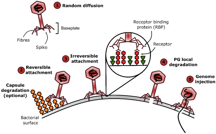
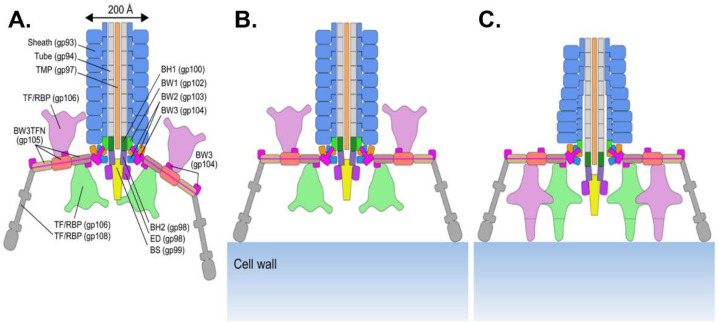
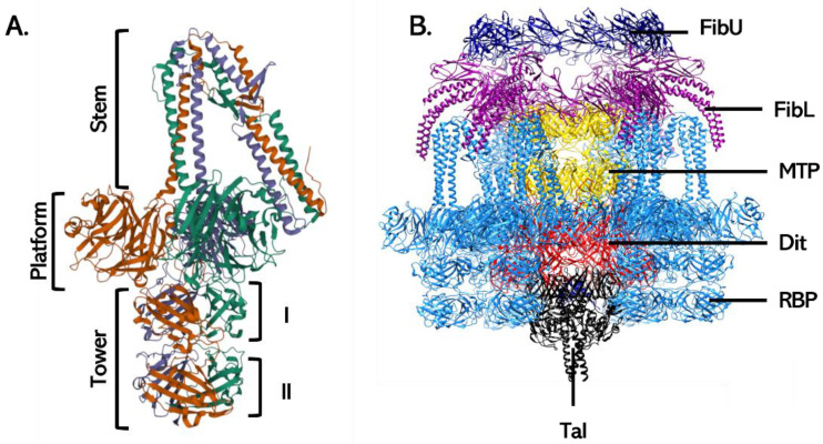
The phage life cycle is a multi-stage process initiated by the recognition and attachment of the virus to its bacterial host. This adsorption step depends on the specific interaction between bacterial structures acting as receptors and viral proteins called Receptor Binding Proteins (RBP). The adsorption process is essential as it is the first determinant of phage host range and a sine qua non condition for the subsequent conduct of the life cycle. In phages belonging to the Caudoviricetes class, the capsid is attached to a tail, which is the central player in the adsorption as it comprises the RBP and accessory proteins facilitating phage binding and cell wall penetration prior to genome injection. The nature of the viral proteins involved in host adhesion not only depends on the phage morphology (i.e., myovirus, siphovirus, or podovirus) but also the targeted host. Here, we give an overview of the adsorption process and compile the available information on the type of receptors that can be recognized and the viral proteins taking part in the process, with the primary focus on phages infecting Gram-positive bacteria.

Keywords: Gram-positive host; adsorption; phage; receptor; receptor binding protein; tail.


Conflict of interest statement

The authors declare no conflict of interest.

Figures

Figure 1
Figure 1
Phage adsorption process and events leading to genome injection. Phage adsorption to their host involves the interaction between Receptor Binding Proteins (RBP) located at the distal part of phage tails and receptors at the surface of the bacterial cell envelope. This process is divided into three steps: (1) random diffusion in the medium, (2) reversible attachment, and (3) irreversible attachment to the bacteria. The adsorption can be assisted by depolymerases cleaving capsular polysaccharides that can hamper receptor access. Following adsorption, virion-associated lysins may locally break down the PG (4) and, finally, conformational changes in the virion structure lead to genome injection (5). This figure was made with the open-source software Inkscape.
Figure 2
Figure 2
Example of secondary cell wall polysaccharides acting as phage receptors. Some SCWP structures are shown on the left with their corresponding SCWP types. The bacterial species in which each SCWP is found are shown on the right, together with one or several examples of phages using these SCWP as receptors. A: d-alanine; P: phosphate; Ac: acetylation. Glycosyl residues: Gal: galactose; GalNAc, N-acetylgalactosamine; Glc: glucose; Gro: glycerol; ManNAc, N-acetylmannosamine; Rbo, ribitol; Rha: rhamnose. SCWP: Secondary Cell Wall Polysaccharide; WTA: Wall Teichoic Acid; LTA: Lipoteichoic Acid; PSP: polysaccharide pellicle; RGP: Rhamnose Glucose polysaccharide. Gray squares indicate unclear linkage units. White hexagons represent PG. In L. lactis PSP are not directly linked to PG but rather to Rhamnose polysaccharides, which are attached to PG. Repeat units are indicated in brackets with numbers representing the number of repeats found in each SCWP, and n referring to an unknown number of repeats. This figure was made with Inkscape, with information from [40,66,70,71,72].
Figure 3
Figure 3
Schematic representation of a myovirus contractile tail. The tail of myoviruses infecting both Gram-positive and Gram-negative hosts harbor conserved proteins that are represented in this scheme. The complete baseplate wedge structure (formed by three types of proteins) is highlighted in red on the right side of the scheme. The table on the right gives the gene product correspondences for each baseplate protein between the two reference phages infecting E. coli (T4 and Mu) and the L. monocytogenes phage A511. TMP: Tape Measure Protein; BW: Baseplate Wedge protein; BH: Baseplate Hub protein; BS: Baseplate Spike; RBP: Receptor Binding Protein; TF: Tail Fiber; MTP: Major Tail Protein.
Figure 4
Figure 4
Examples of baseplate organizations found in siphoviruses. (A) Tail proteins are usually encoded between the TMP and the lysis cassette (not shown here). Gene identification is indicated in each arrow. TMP are shown in gray, Dit in yellow, Tal in green, RBP in red, other tail proteins in blue, and hypothetical proteins in white. (B) Schematic representation of p2, TP901-1, SPP1, λ, and T5 baseplates recognizing either carbohydrate or protein receptors. Color coding is that of the panel (A). TMP: Tape Measure Protein; Dit: Distal tail; Tal: Tail lysin; LTF: Long Tail Fiber; BppU: Upper Baseplate protein. This figure was made with Inkscape.
Figure 5
Figure 5
Structure of Dit proteins of L. lactis phage p2 (left) and B. subtilis phage SPP1 (right). Dit proteins form hexameric structures, and one monomer is highlighted in red. In p2 Dit protein, the “arm” structure is indicated by an arrow. Structures were retrieved from the Protein Data Bank (p2 Dit: PDB ID 4V5I and [119]; SPP1 Dit: PDB ID 2X8K and [118]).
Figure 6
Figure 6
Baseplate conformation of siphoviruses. (A) Phage TP901-1 has a “ready-to-infect” baseplate, which is formed by a Dit hexamer (red) on which are anchored six trimeric BppU (green), each attaching three RBP trimer (blue). One tripod is formed by one BppU attaching three RBP and is highlighted in a different shade of black. The Tal trimer attaching at the bottom of the baseplate is missing. (B) In presence of divalent cation, phage p2 baseplate switches from a “rest” conformation, where RBP point upwards toward the capsid, to an “active” conformation where the RBP face down. The trimeric Tal (black) and the six RBP trimers (blue) are attached to a hexameric Dit (dark red). In phage p2, a second Dit hexamer is found in the baseplate (pink). Structures were retrieved from the Protein Data Bank (TP901-1 baseplate: PDB ID 4V96 and [91]; p2 baseplate rest: PDB ID 6ZJJ and [120]; p2 baseplate active: PDB ID 6ZIH and [120]).
Figure 7
Figure 7
Schematic representation of podovirus Φ29 infecting B. subtilis. Gp numbers indicate the proteins involved in such structures in phage phi29. This figure was made with Inkscape.
Figure 8
Figure 8
Structure of lactococcal RBP. Most lactococcal RBP harbor a three-domain organization with an N-terminal (Nt) shoulder domain, which attaches the RBP to the virion, a central connecting neck domain, and a C-terminal (Ct) head domain involved in receptor recognition. In some phages (e.g., 1358), the neck domain is absent. Each protein monomer is displayed in a different color. Structures were retrieved from the Protein Data Bank (1358: PDB ID 4RGA and [152]; p2: PDB ID 2BSD and [153]; TP901-1: PDB ID 3EJC and [134]).
Figure 9
Figure 9
Listeria phage A511 baseplate structure and baseplate transformation upon adsorption. (A) Phage A511 tail structure. The name of each tail protein is indicated with the related gene in parentheses. (B) A511 first interaction with the CW through binding of the RBP gp108. (C) Reorientation of the gp106 pyramidal structures and interaction with the CW. The distal part of the sheath begins to contract. BH: Baseplate Hub; RBP: Receptor Binding Protein; BW: Baseplate Wedge; BS: Baseplate Spike; TF: Tail fiber; TFN: Tail Fiber Network. Reprinted with permission from [99].
Figure 10
Figure 10
Tail proteins constituting the baseplate of S. aureus siphoviruses. (A) The RBP of ϕ11 (gp45) has a modular organization with three domains. N-terminal stem domain, central platform with the receptor binding site, and C-terminal tower domain consisting of two similar regions. (B) Phage 80α baseplate is particular in that it harbors, in addition to the RBP, two other tail fibers (FibL and FibU) that may be involved in adsorption. The Tal and FibU could not be completely modeled, and only the N-terminal part of the FibL are shown for clarity purposes. FibU: Upper tail fiber (dark blue); FibL: Lower tail fiber (purple); RBP: Receptor Binding Protein (blue); Tal: Tail lysin (black). MTP: Major Tail Protein (Yellow). Structures were retrieved from the Protein Data Bank (ϕ11 RBP: PDB 5EFV and [129]; 80α baseplate: 6V8I and [128]).

References

    1. Dion M.B., Oechslin F., Moineau S. Phage diversity, genomics and phylogeny. Nat. Rev. Microbiol. 2020;18:125–138. doi: 10.1038/s41579-019-0311-5. - DOI - PubMed
    1. Bertozzi Silva J., Storms Z., Sauvageau D. Host receptors for bacteriophage adsorption. FEMS Microbiol. Lett. 2016;363:fnw002. doi: 10.1093/femsle/fnw002. - DOI - PubMed
    1. Nobrega F.L., Vlot M., de Jonge P.A., Dreesens L.L., Beaumont H.J.E., Lavigne R., Dutilh B.E., Brouns S.J.J. Targeting mechanisms of tailed bacteriophages. Nat. Rev. Microbiol. 2018;16:760–773. doi: 10.1038/s41579-018-0070-8. - DOI - PubMed
    1. Munsch-Alatossava P., Alatossava T. The extracellular phage-host interactions involved in the bacteriophage ll-h infection of Lactobacillus Delbrueckii sp. Lactis ATCC 15808. Front. Microbiol. 2013;4:408. doi: 10.3389/fmicb.2013.00408. - DOI - PMC - PubMed
    1. Degroux S., Effantin G., Linares R., Schoehn G., Breyton C. Deciphering bacteriophage T5 host recognition mechanism and infection trigger. BioRxiv. 2022 doi: 10.1101/2022.09.22.509047. - DOI - PMC - PubMed

Publication types

LinkOut - more resources