. 2023 Jan 10;15(1):202.
doi: 10.3390/v15010202.

Development of HIV-Resistant CAR T Cells by CRISPR/Cas-Mediated CAR Integration into the CCR5 Locus

Affiliations

Development of HIV-Resistant CAR T Cells by CRISPR/Cas-Mediated CAR Integration into the CCR5 Locus

Frederik Holm Rothemejer et al. Viruses. .

Abstract

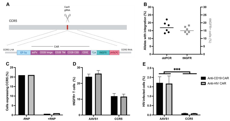
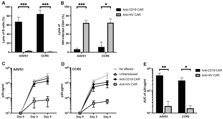
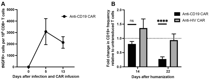
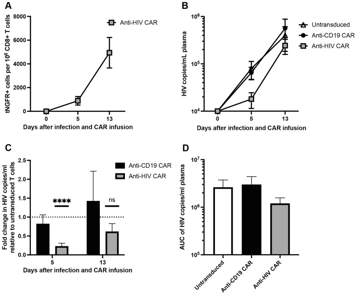
Adoptive immunotherapy using chimeric antigen receptor (CAR) T cells has been highly successful in treating B cell malignancies and holds great potential as a curative strategy for HIV infection. Recent advances in the use of anti-HIV broadly neutralizing antibodies (bNAbs) have provided vital information for optimal antigen targeting of CAR T cells. However, CD4+ CAR T cells are susceptible to HIV infection, limiting their therapeutic potential. In the current study, we engineered HIV-resistant CAR T cells using CRISPR/Cas9-mediated integration of a CAR cassette into the CCR5 locus. We used a single chain variable fragment (scFv) of the clinically potent bNAb 10-1074 as the antigen-targeting domain in our anti-HIV CAR T cells. Our anti-HIV CAR T cells showed specific lysis of HIV-infected cells in vitro. In a PBMC humanized mouse model of HIV infection, the anti-HIV CAR T cells expanded and transiently limited HIV infection. In conclusion, this study provides proof-of-concept for developing HIV-resistant CAR T cells using CRISPR/Cas9 targeted integration.

Keywords: CAR T cells; CRISPR/Cas9; HIV; humanized mice.


Conflict of interest statement

The authors declare no conflict of interest.

Figures

Figure 1
Figure 1
CRISPR-Cas9 integration of a CAR into CCR5 leads to an HIV-resistant phenotype: (A) schematic of the CAR construct; (B) comparison of CAR genes specifically integrated into CCR5 by ddPCR seven days after transduction (left axis) and tNGFR expression by flow cytometry four days after transduction (right axis); (C) CCR5 expression by flow cytometry in cells treated with or without CCR5-sgRNA/Cas9 RNP and transduced with CCR5-CAR AAV6; (D) integration rates of either anti-CD19 CAR or anti-HIV CAR into either AAVS1 or CCR5; (E) levels of eGFP expression in CD4+ CAR T cells 24 h after infection with eGFP-HIV (data presented as means ± SEM, ***: p ≤ 0.001).
Figure 2
Figure 2
Anti-HIV CAR T cells specifically kill HIV-infected cells in vitro: (A) cytotoxicity assay of autologous B cells at E:T 5:1; (B) cytotoxicity assay of HIV-infected autologous CD4+ T cells at E:T 1:1; (C,D) levels of p24 in supernatants following five day co-culture of CAR T cells and HIV-infected CD8-depleted autologous PBMCs at E:T 1:1. (C): CARs are integrated into AAVS1, (D) CARs are integrated into CCR5; (E) AUC of p24 curves in (C,D) (data presented as means ± SEM, *: p ≤ 0.05, **: p ≤ 0.01, ***: p ≤ 0.001).
Figure 3
Figure 3
Anti-CD19 CAR T cells kill B cells in vivo: (A) frequency of anti-CD19 CAR T cells based on the frequency of tNGFR+ cells per 1 × 10⁶ CD8+ T cells; (B) fold change in the frequency of CD19+ cells for each CAR T cell-treated group normalized to the frequency of CD19+ cells in mice treated with untransduced CD8+ T cells (n = 7–8 mice from 2 biological donors in each group, data presented as means ± SEM, ns: p > 0.05, ****: p ≤ 0.0001).
Figure 4
Figure 4
Anti-HIV CAR T cells expand and transiently limit HIV infection in vivo: (A) frequency of anti-HIV CAR T cells based on the frequency of tNGFR+ cells per 1 × 10⁶ CD8+ T cells; (B) HIV viral load as copies/mL plasma; (C) fold change in viral load for each CAR T cell-treated group normalized to the viral load in mice treated with untransduced CD8+ T cells; (D) AUC of viral load in (B) (n = 7–8 mice from 2 biological donors in each group, data presented as means ± SEM, ns: p > 0.05, ****: p ≤ 0.0001).

References

    1. Schuster S.J., Svoboda J., Chong E.A., Nasta S.D., Mato A.R., Anak O., Brogdon J.L., Pruteanu-Malinici I., Bhoj V., Landsburg D., et al. Chimeric Antigen Receptor T Cells in Refractory B-Cell Lymphomas. N. Engl. J. Med. 2017;377:2545–2554. doi: 10.1056/NEJMoa1708566. - DOI - PMC - PubMed
    1. Park J.H., Riviere I., Gonen M., Wang X., Senechal B., Curran K.J., Sauter C., Wang Y., Santomasso B., Mead E., et al. Long-Term Follow-up of CD19 CAR Therapy in Acute Lymphoblastic Leukemia. N. Engl. J. Med. 2018;378:449–459. doi: 10.1056/NEJMoa1709919. - DOI - PMC - PubMed
    1. Munshi N.C., Anderson L.D., Jr., Shah N., Madduri D., Berdeja J., Lonial S., Raje N., Lin Y., Siegel D., Oriol A., et al. Idecabtagene Vicleucel in Relapsed and Refractory Multiple Myeloma. N. Engl. J. Med. 2021;384:705–716. doi: 10.1056/NEJMoa2024850. - DOI - PubMed
    1. Kim G.B., Hege K., Riley J.L. CAR Talk: How Cancer-Specific CAR T Cells Can Instruct How to Build CAR T Cells to Cure HIV. Front. Immunol. 2019;10:2310. doi: 10.3389/fimmu.2019.02310. - DOI - PMC - PubMed
    1. Mitsuyasu R.T., Anton P.A., Deeks S.G., Scadden D.T., Connick E., Downs M.T., Bakker A., Roberts M.R., June C.H., Jalali S., et al. Prolonged survival and tissue trafficking following adoptive transfer of CD4ζ gene-modified autologous CD4+ and CD8+ T cells in human immunodeficiency virus–infected subjects. Blood. 2000;96:785–793. doi: 10.1182/blood.V96.3.785.015k10_785_793. - DOI - PubMed

Publication types