. 2023 Apr;65(4):835-843.
doi: 10.1007/s00234-022-03111-8. Epub 2023 Jan 21.

Assessment of the levels of termination of the conus medullaris and thecal sac in the pediatric population

Affiliations

Assessment of the levels of termination of the conus medullaris and thecal sac in the pediatric population

Mohammed Blaaza et al. Neuroradiology. 2023 Apr.

Abstract

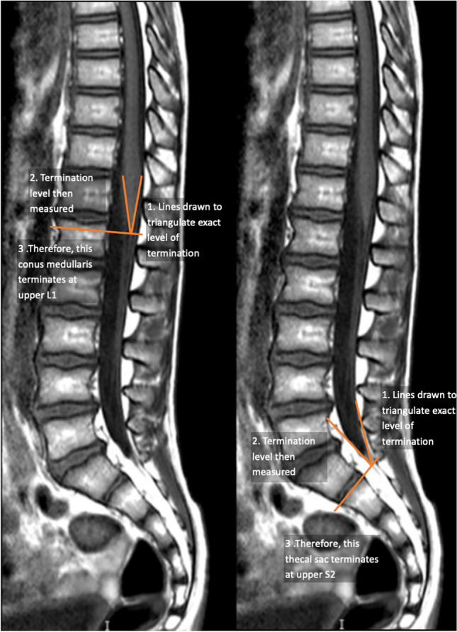
Purpose: This study assessed the position of the termination of the conus medullaris (the point where the spinal cord tapers to an end) and thecal sac (the sheath of dura mater that surrounds the spinal cord and caudal nerve roots) in a large pediatric population, to characterise the nature of the pediatric Gaussian distribution and assess whether age affected the distribution. The study further aimed to assess the effect of gender on termination positions.

Methods: A total of 520 MRI spine studies of children aged between 1 month and 19 years old were collected from two pediatric tertiary referral centres in the UK and Italy. Studies with pathological findings were excluded, and normal scans were found using keyword search algorithms on a database of radiologists' reports. The reported scans were individually assessed and reviewed by two experienced neuroradiologists. The termination points of the conus medullaris and thecal sac were determined for each study. Local IRB approvals were sought.

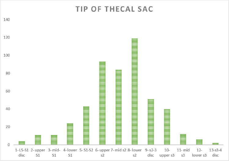
Results: The results showcased a Gaussian distribution in both conus medullaris (r=0.8997) and thecal sac termination levels (r=0.9639). No statistically significant results were noted with increasing age for the termination positions of the conus medullaris or thecal sac (p = 0.154, 0.063). No statistical significance was observed with gender variation with either anatomical landmark. A weak positive correlation was observed between the termination levels of the conus medullaris and the thecal sac (r=0.2567) CONCLUSION: Termination levels across all pediatric age range followed a Gaussian distribution. Knowledge of normal termination levels has relevant clinical implications, including the assessment of patients with suspected spinal dysraphism.

Keywords: Conus medullaris; Conus termination; Pediatric spine; Thecal sac.


Conflict of interest statement

None of the authors have any conflicts of interest to disclose.

Figures

Fig. 1
Fig. 1
A showcase of how the level of termination of the thecal sac and the conus medullaris were measured in steps 1–3
Fig. 2
Fig. 2
Graph showing the distribution of conus medullaris termination levels in the patient population. Termination position is shown on the x-axis, and the number of studies terminating at each level is shown on the y-axis
Fig. 3
Fig. 3
Graph showing the distribution of termination levels of the conus medullaris in male and female pediatric patients. Termination position is shown on the x-axis, and the number of studies terminating at each level is shown on the y-axis
Fig. 4
Fig. 4
Graph showing the distribution of thecal sac termination levels in the patient population. Termination position is shown on the x-axis, and the number of studies terminating at each level is shown on the y-axis
Fig. 5
Fig. 5
Graph showing the distribution of termination levels of the thecal sac in male and female pediatric patients. Termination position is shown on the x-axis, and the number of studies terminating at each level is shown on the y-axis
Fig. 6
Fig. 6
Graph showing the correlation between the levels of termination of the conus medullaris and thecal sac. Termination levels of conus medullaris are shown on the x-axis and that of the thecal sac on the y-axis
Fig. 7
Fig. 7
Graph showing the effect of age on the distributions of termination level of the conus medullaris in the first year of life. Age in months is shown on the x-axis, and the termination position is shown on the y-axis.
Fig. 8
Fig. 8
Graph showing the effect of age on the distributions of termination level of the thecal sac in the first year of life. Age in months is shown on the x-axis, and termination position is shown on the y-axis

References

    1. Nene Y, Jilani TN. StatPearls. Treasure Island (FL): StatPearls Publishing; 2022. Neuroanatomy, Conus Medullaris. - PubMed
    1. Kesler H, Dias M, Kalapos P. Termination of the normal conus medullaris in children: a whole-spine magnetic resonance imaging study. Neurosurgical Focus. 2007;23(2):1–5. doi: 10.3171/FOC-07/08/E7. - DOI - PubMed
    1. Jung J, Kim E, Song I, Lee J, Kim H, Kim J. The influence of age on positions of the conus medullaris, Tuffier’s line, dural sac, and sacrococcygeal membrane in infants, children, adolescents, and young adults. Pediatric Anesthesia. 2016;26(12):1172–1178. doi: 10.1111/pan.12998. - DOI - PubMed
    1. Wilson D, Prince J MR imaging determination of the location of the normal conus medullaris throughout childhood. 10.1007/978-3-662-11158-1_89 - PubMed
    1. Asil K et al (2018) Conus medullaris levels on ultrasonography in term newborns: normal levels and dermatological findings. 10.3340/jkns.2016.1212.001 - PMC - PubMed