. 2023 Apr 1:225:115064.
doi: 10.1016/j.bios.2023.115064. Epub 2023 Jan 5.

Spatiotemporal dissection of tumor microenvironment via in situ sensing and monitoring in tumor-on-a-chip

Affiliations

Spatiotemporal dissection of tumor microenvironment via in situ sensing and monitoring in tumor-on-a-chip

Lang Zhou et al. Biosens Bioelectron. .

Abstract

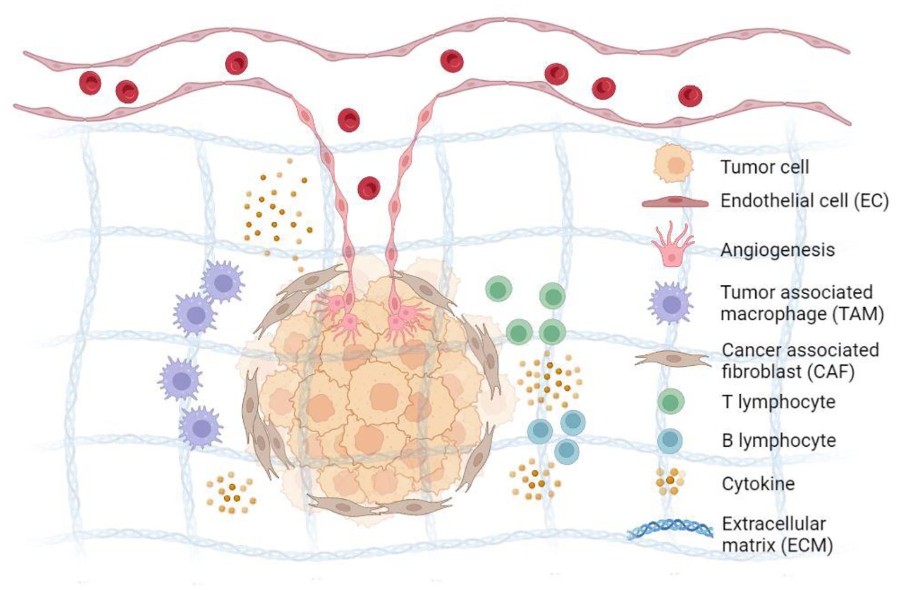
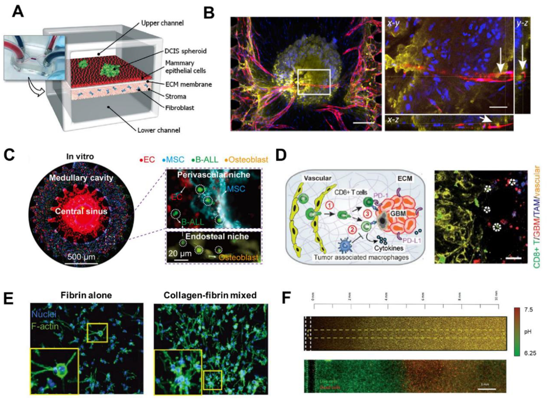
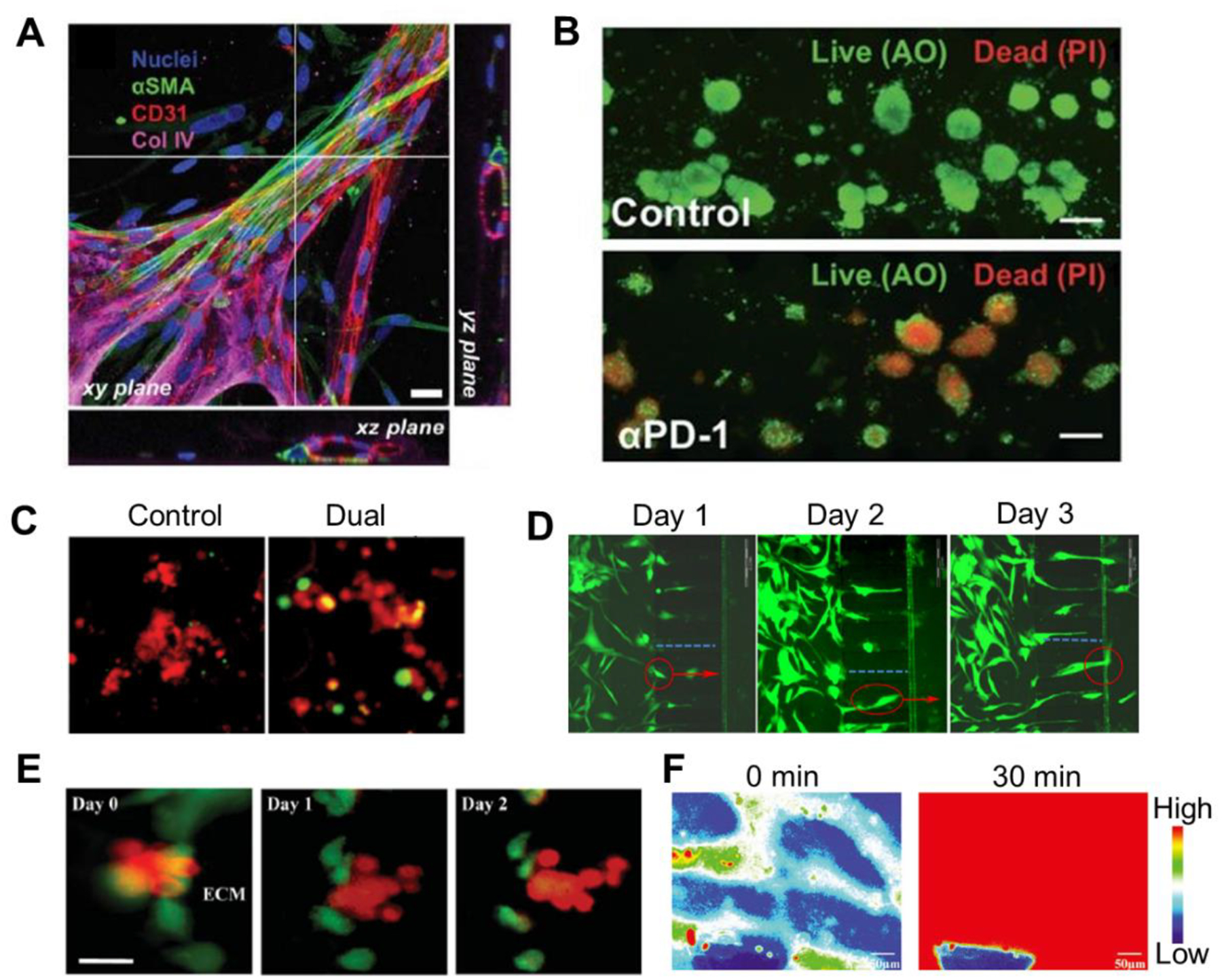
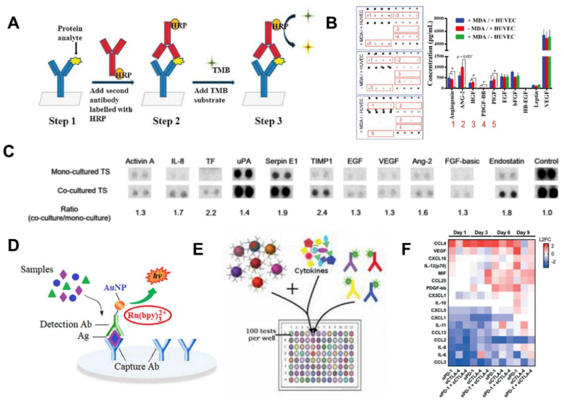
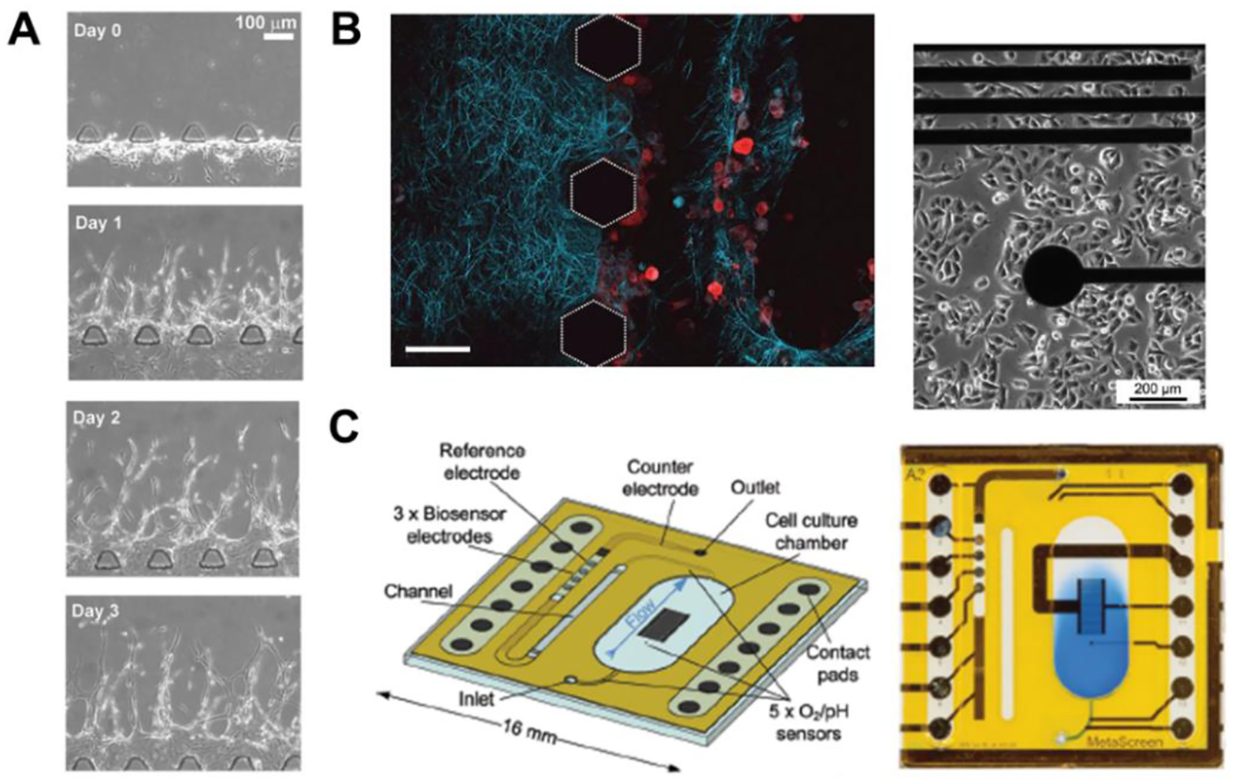
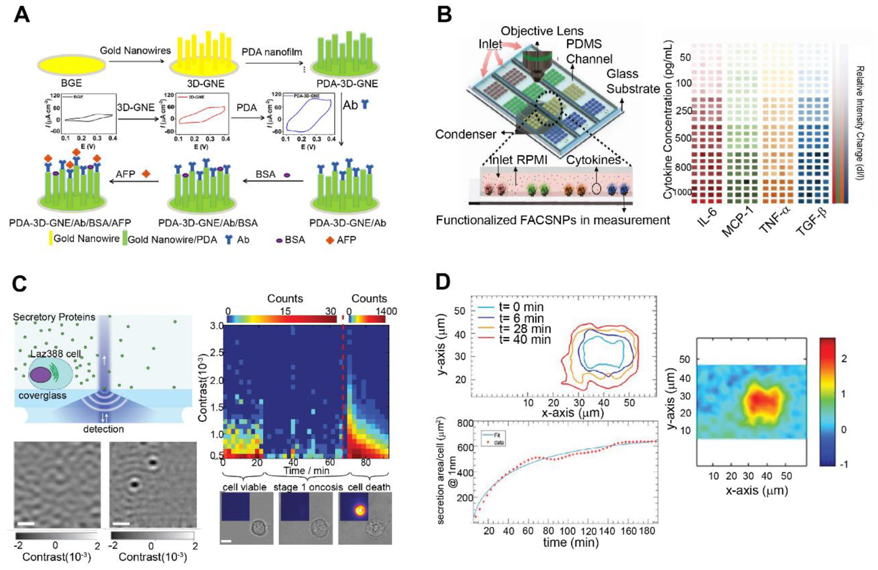
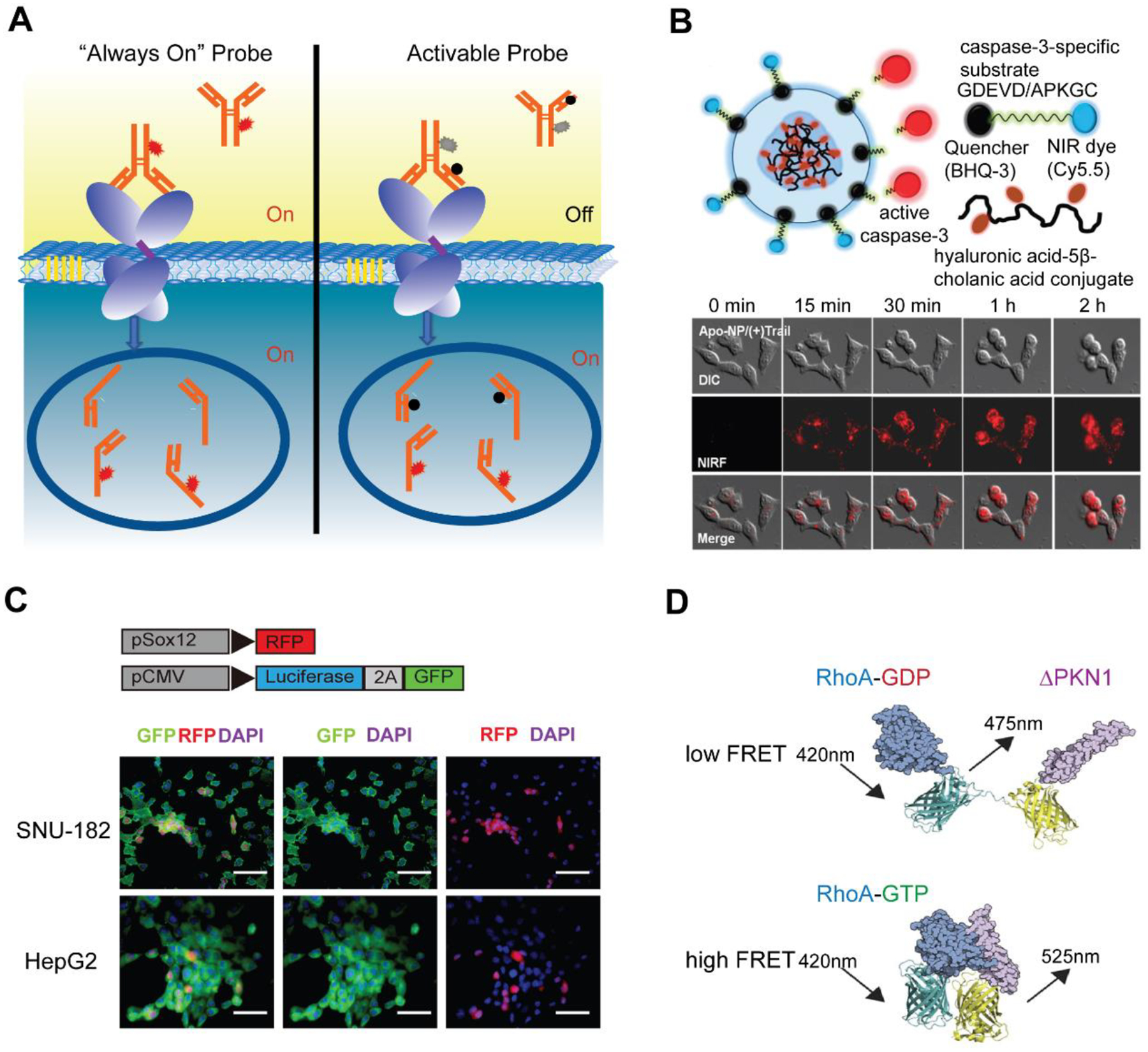
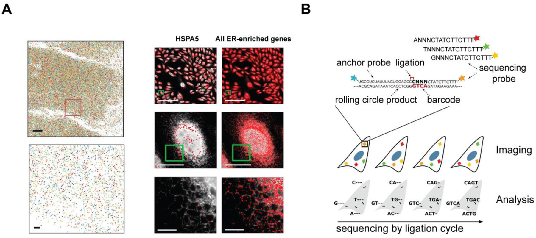
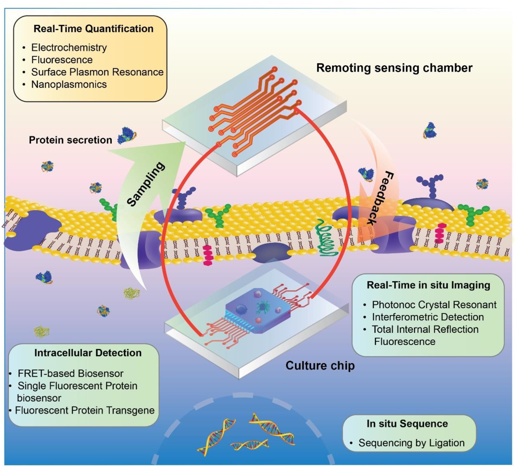
Real-time monitoring in the tumor microenvironment provides critical insights of cancer progression and mechanistic understanding of responses to cancer treatments. However, clinical challenges and significant questions remain regarding assessment of limited clinical tissue samples, establishment of validated, controllable pre-clinical cancer models, monitoring of static versus dynamic markers, and the translation of insights gained from in vitro tumor microenvironments to systematic investigation and understanding in clinical practice. State-of-art tumor-on-a-chip strategies will be reviewed herein, and emerging real-time sensing and monitoring platforms for on-chip analysis of tumor microenvironment will also be examined. The integration of the sensors with tumor-on-a-chip platforms to provide spatiotemporal information of the tumor microenvironment and the associated challenges will be further evaluated. Though optimal integrated systems for in situ monitoring are still in evolution, great promises lie ahead that will open new paradigm for rapid, comprehensive analysis of cancer development and assist clinicians with powerful tools to guide the diagnosis, prognosis and treatment course in cancer.

Keywords: In situ biosensing; Integrated system; Real-time monitoring; Spatiotemporal analysis; Tumor microenvironment; Tumor-on-a-chip.


Conflict of interest statement

Declaration of competing interest The authors declare that they have no known competing financial interests or personal relationships that could have appeared to influence the work reported in this paper.

Figures

Figure 1.
Figure 1.. Components of the tumor microenvironment.
Tumor microenvironment consists of multiple cells including tumor cells, endothelial cells forming vasculature participating in angiogenesis and metastasis, cancer associated fibroblasts, tumor associated macrophages, and lymphocytes. Cytokines and chemokines released by malignant and stromal cells inhibit the function of resident immune cells, leading to the TME immunosuppression. Extracellular matrices facilitate invasion of tumor cells through matrix remodeling. Metabolic factors like pH and glucose are suppressed in tumor milieu. Lastly, hypoxia, the lack of oxygen, also plays a key role in the evolution of tumors, contributing to tumor invasion and metastasis.
Figure 2.
Figure 2.. Modeling TME on tumor-on-a-chip with multiple components.
(A) A human breast-cancer-on-a-chip replicating the microarchitecture of breast ductal carcinoma with integration of tumor spheroids, mammary ductal epithelial cells, and mammary fibroblasts. Adapted with permission. Copyright 2015, the Royal Society of Chemistry. (Choi et al. 2015). (B) Immunofluorescence images of the tumor spheroid integrated with the blood vessels on a vascularized TOC. Lumen structure of vasculatures was observed, indicating the perfusability of vascular network. Adapted with permission. Copyright 2020, Elsevier. (Nashimoto et al. 2020). (C) Whole scan of the leukemic BM niche system. Compartments on chip resembled the in vivo counterparts like central sinus and medullary cavity with niche cells. Adapted under the terms of the CC-BY-NC license. Copyright 2020, the Authors. (Ma et al. 2020) (D) Immunosuppressive TME on GBM-on-a-chip. The reconstituted GBM TME recapitulated the infiltration of T cells and interactions among T cells, TAMs, and GBM tumor cells. Adapted under the terms of the CC-BY license. Copyright 2020, the Authors. (Cui et al. 2020) (E) Various components of ECM loaded on TOC revealed the distinct phenotypic changes of cancer cells in response to different ECM conditions. Adapted with permission. Copyright 2017, Wiley. (Chung et al. 2017). (F) pH gradient and necrotic region were formed on TOC under metabolic starvation gradients. Adapted with permission. Copyright 2019, the Royal Society of Chemistry. (Ayuso et al. 2019).
Figure 3.
Figure 3.. Fluorescence-based methods.
(A) Immunostaining image of a pericyte-decorated blood vessel: microvascular network (CD31, red) covered with pericytes (a-SMA, green) Adapted with permission. Copyright 2013, the Royal Society of Chemistry. (Kim et al. 2013) (B) Live/Dead imaging of organotypic tumor spheroids with acridine orange (AO) and propidium iodide (PI) labelling. Adapted with permission. Copyright 2018, the Royal Society of Chemistry. (Aref et al. 2018) (C) Increased apoptosis of tumor cells when receiving combinational immunotherapy was shown by caspase-3/7 activation. Adapted under the terms of the CC-BY license. Copyright 2020, the Authors. (Cui et al. 2020). (D) Temporal images of GFP-labeled cancer cell migration, where red circle indicated the same cancer cell in different time points. Adapted under the terms of the CC-BY license. Copyright 2016, the Authors. (Mi et al. 2016) (E) Transendothelial invasion of tumor aggregates induced by CXCL12, where cancer cells were labeled with CellTracker Red (red) and endothelial cells were labeled with CellTracker Green (green). Adapted with permission. Copyright 2012, the Royal Society of Chemistry. (Zhang et al. 2012). (F) Heat map of fluorescence intensity denotes diffusion of FITC-dextran from a more permeable vascular network due to the presence of tumor cells. Adapted with permission. Copyright 2018, Wiley. (Nagaraju et al. 2018).
Figure 4.
Figure 4.. ELISA-based analysis.
(A) Concepts of ELISA. Adapted with permission. Copyright 2014, the Royal Society of Chemistry. (Zhang et al. 2014) (B) Profiling ten angiogenesis-related cytokines in the tumor-vascular TME on chip with multiplex ELISA. Adapted with permission. Copyright 2018, Wiley. (Nagaraju et al. 2018) (C) Chemiluminescence-based proteome array analysis of angiogenesis and metastasis-related cytokines in tumor spheroids-stellate cells co-culture system. 11 factors among 55 tested cytokines showed significant statistic differences. Adapted under the terms of the CC-BY license. Copyright 2018, the Authors. (Lee et al. 2018a) (D) Concepts of a sandwich-based ECLIA using gold nanoparticles as both label and co-reactant. Adapted with permission. Copyright 2020, Elsevier. (Zhao et al. 2020a) (E) Principle of microbead-based sandwich immunoassay. Plastic beads with red and infrared fluorescent dyes provide unique signatures to samples while the detection antibodies conjugated with a reporter dye are added to measure cytokines. Adapted with permission. Copyright 2015, Elsevier. (Khalifian et al. 2015). (F) Time-lapse heatmap of secretory cytokines under different immune checkpoint blockade treatments from tumor spheroids on a chip measured by bead-based immunoassay. Adapted with permission. Copyright 2018, the Royal Society of Chemistry. (Aref et al. 2018).
Figure 5.
Figure 5.. Other current analysis methods.
(A) Phase contrast microphotographs of migration of endothelial cells towards leukemia cells over time on a biomimetic angiogenesis chip. Adapted with permission. Copyright 2017, Wiley. (Zheng et al. 2016) (B) The degradation of collagen fibrils (in blue) caused by the migration of breast cancer cells (in red) was imaged via SHG within a microfluidic device. Adapted with permission. Copyright 2009, the Royal Society of Chemistry. (Huang et al. 2009). (C) Metabolic factors sensing chip by integrating O2, pH, glucose and lactate biosensors in one microfluidic device. Adapted with permission. Copyright 2014, the Royal Society of Chemistry. (Weltin et al. 2014)
Figure 6.
Figure 6.. Secretory protein analysis via real-time detection biosensors.
(A) Nanoelectrodes for detection of α-fetoprotein with a detection limit of 0.01 pg/mL. Adapted with permission. Copyright 2020, Elsevier. (Hong et al. 2020) (B) multiarrayed LSPR optical immunoassay based on a magnet patterned Fe3O4/Au core–shell nanoparticle sensing array for simultaneous real-time detection of four cytokines in complex biological samples, with limit of detection at ~20 pg/mL. Adapted with permission. Copyright 2019, Wiley. (Cai et al. 2019) (C) Interferometric detection of scattered light (iSCAT) method for in-situ real-time imaging of secreted proteins by Epstein−Barr virus -transformed B cell line. Reprinted adapted with permission from (McDonald et al. 2018). Copyright 2018, American Chemical Society. (D) Photonic crystal resonant (PCR) imaging of thrombopoientin secretion at single-cell level, with fine resolution of 2~6 μm and detection limit below 125 ng/mL. Adapted under the terms of the CC-BY license. Copyright 2018, the Authors. (Juan-Colaás et al. 2018)
Figure 7.
Figure 7.. Fluorescence Molecular Imaging for dynamic biomarker analysis.
(A) Principles of “always on” and activatable probe for biomarker recognition. (B) FRET based activatable probes (dual-quenched caspase-3-sensitive peptides) used for monitoring caspase-3 activity. Reprinted adapted with permission from (Lee et al. 2011). Copyright 2011, American Chemical Society. (C) Reporter red fluorescence protein (RFP) gene fused with a Sox12 promoter for the study of Sox12 as a potential cancer stem-like Cell marker in Hepatocellular Carcinoma. Adapted under the terms of the CC-BY license. Copyright 2017, the Authors. (Zou et al. 2017) (D) FRET-based FP biosensor used for activity imaging of plasma membrane restricted small GTPase RhoA. Adapted under the terms of the CC-BY license. Copyright 2015, the Authors. (Van Unen et al. 2015)
Figure 8.
Figure 8.. In situ hybridization/sequencing for TME validation
(A) Multiplexed error-robust fluorescence in situ hybridization (MERFISH) for 10,050-gene imaging in individual U-2 OS cells. Left: U-2 OS sample stained with encoding probes targeting 10,050 RNA species and a zoomed-in image below (Scale bar: 1 μm.). Right: identification of RNAs enriched at the endoplasmic reticulum. Adapted under the terms of the CC BY-NC-ND license. Copyright 2020, the Authors. (Xia et al. 2019) (B) Schematic for in situ sequencing of barcoded mRNA amplicons in fixed cells. Adapted under the terms of the CC-BY license. Copyright 2019, the Authors. (Maïno et al. 2019)
Figure 9.
Figure 9.. Conceptual illustration of integrated TOC systems.
The integration of viable techniques for real-time sensing and monitoring of TME in TOCs.

Similar articles

Cited by

References

    1. Abbott J, Ye T, Krenek K, Gertner RS, Ban S, Kim Y, Qin L, Wu W, Park H, Ham D, 2020. Nat. Biomed. Eng 4(2), 232–241. - PMC - PubMed
    1. Ahn J, Sei YJ, Jeon NL, Kim Y, 2017. Bioengineering 4(3). - PMC - PubMed
    1. Albanese A, Lam AK, Sykes EA, Rocheleau JV, Chan WCW, 2013. Nat. Commun 4(1), 2718. - PMC - PubMed
    1. Anderson NM, Simon MC, 2020. Curr. Biol 30(16), R921–R925. - PMC - PubMed
    1. Ando Y, Ta HP, Yen DP, Lee S-S, Raola S, Shen K, 2017. Sci. Rep 7(1), 15233. - PMC - PubMed