. 2023 Apr:60:102612.
doi: 10.1016/j.redox.2023.102612. Epub 2023 Jan 13.

Red blood cells from endothelial nitric oxide synthase-deficient mice induce vascular dysfunction involving oxidative stress and endothelial arginase I

Affiliations

Red blood cells from endothelial nitric oxide synthase-deficient mice induce vascular dysfunction involving oxidative stress and endothelial arginase I

Zhengbing Zhuge et al. Redox Biol. 2023 Apr.

Abstract

Background & aims: Nitric oxide bioactivity (NO) from endothelial NO synthase (eNOS) importantly contributes to the maintenance of vascular homeostasis, and reduced eNOS activity has been associated with cardiovascular disease. Emerging evidence suggests interaction(s) between red blood cells (RBCs) and the endothelium in vascular control; however, the specific role of RBC eNOS is less clear. We aimed to investigate the hypothesis that a lack of RBC eNOS induces endothelial dysfunction.

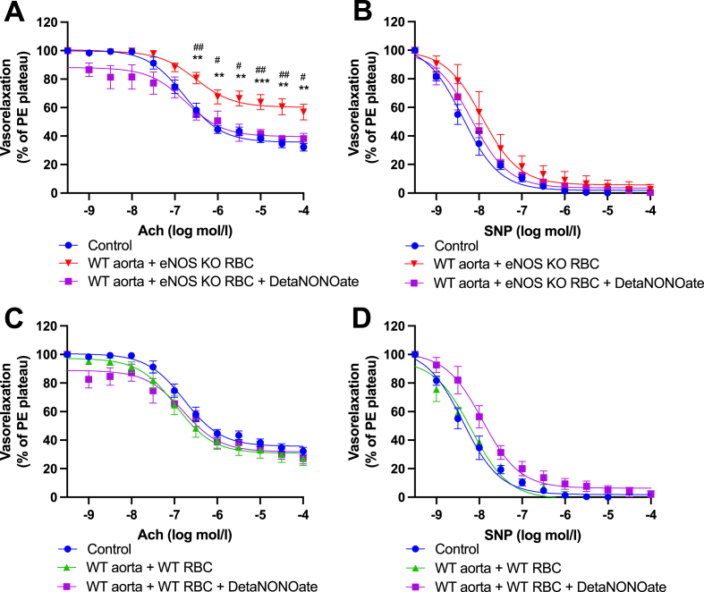
Methods & results: RBCs from global eNOS knockout (KO) and wildtype (WT) mice were co-incubated ex vivo overnight with healthy mouse aortic rings, followed by functional and mechanistic analyses of endothelium-dependent and independent relaxations. RBCs from eNOS KO mice induced endothelial dysfunction and vascular oxidative stress, whereas WT RBC did not. No differences were observed for endothelium-independent relaxations. This eNOS KO RBC-induced endothelial dysfunctional phenotype was prevented by concomitant co-incubation with reactive oxygen species scavenger (TEMPOL), arginase inhibitor (nor-NOHA), NO donor (detaNONOate) and NADPH oxidase 4 (NOX4) inhibitor. Moreover, vessels from endothelial cell-specific arginase 1 KO mice were resistant to eNOS KO-RBC-induced endothelial dysfunction. Finally, in mice aortae co-incubated with RBCs from women with preeclampsia, we observed a significant reduction in endothelial function compared to when using RBCs from healthy pregnant women or from women with uncomplicated gestational hypertension.

Conclusions: RBCs from mice lacking eNOS, and patients with preeclampsia, induce endothelial dysfunction in adjacent blood vessels. Thus, RBC-derived NO bioactivity acts to prevent induction of vascular oxidative stress occurring via RBC NOX4-derived ROS in a vascular arginase-dependent manner. Our data highlight the intrinsic protective role of RBC-derived NO bioactivity in preventing the damaging potential of RBCs. This provides novel insight into the functional relationship between RBCs and the vasculature in health and cardiovascular disease, including preeclampsia.

Keywords: Arginase; Nitric oxide; Oxidative stress; Red blood cells; eNOS.


Conflict of interest statement

Declaration of competing interest The authors declare that they have no known competing financial interests or personal relationships that could have appeared to influence the work reported in this paper.

Figures

Image 1
Graphical abstract
Fig. 1
Fig. 1
Overview of experimental protocol. Aortae and whole blood were isolated from eNOS WT and global eNOS KO mice. Whole blood constituents were separated, and red blood cells (RBC) were washed. Aortae were cleaned and separated into aortic rings in PSS. In indicated combinations, aortic rings and washed RBCs (10% haematocrit) were co-incubated (37 °C; 5% CO2) overnight (18 h). Following incubation, aortic rings were washed, and vessel reactivity (vasorelaxation and contractility) was assessed via vascular wire myography. To further assess the role of reactive oxygen species (ROS), arginase and NADPH oxidase (NOX) activity on the observed functional interaction between eNOS KO RBCs and WT aortae, the co-incubations were simultaneously treated with TEMPOL, nor-NOHA and NOX inhibitors, respectively.
Fig. 2
Fig. 2
RBCs from eNOS KO mice induce endothelial dysfunction. (A) Endothelial-dependent vasorelaxation (% of Phenylephrine plateau; PE) response curve to acetylcholine (Ach; log mol/L) and (B) endothelial-independent vasorelaxation (% of phenylephrine plateau; PE) response curve to nitroprusside (SNP) of mouse aortic rings following overnight incubation with Control, RBC from a WT mouse (WT RBC) or RBC from an eNOS KO mouse (eNOS KO RBC). Control and eNOS KO aorta incubation denotes incubation of a WT or eNOS KO aorta, respectively, with DMEM media without RBC. Data expressed as mean ± SEM; Control, n = 7; WT aorta + WT RBC, n = 17; WT aorta + eNOS KO RBC, n = 18; eNOS KO aorta; n = 10, analysed via ordinary 2-way ANOVA with multiple comparisons and Tukey’s post-hoc test. Statistical significance defined as *p < 0.05, **p < 0.01; ***p < 0.005; ****p < 0.001; Comparisons of WT aorta + eNOS KO RBC *vs Control; # vs WT aorta + WT RBC; † vs eNOS KO aorta.
Fig. 3
Fig. 3
Heme-NO signal is not detected in RBCs from eNOS KO mice. (A) EPR spectra and (B) quantification of Heme-NO signal in mouse RBCs. Spectra were recorded at 77K. Instrument settings indicated in methods. Analysed via a Students t-test for unpaired observations. Statistical significance defined as * p < 0.05, ***p < 0.005.
Fig. 4
Fig. 4
Functional role of dysregulated NO homeostasis in eNOS KO RBC-induced endothelial dysfunction. (A, C) Endothelial-dependent vasorelaxation (% of Phenylephrine plateau; PE) response curve to acetylcholine (Ach; log mol/L) and (B, D) endothelial-independent vasorelaxation (% of phenylephrine plateau; PE) response curve to nitroprusside (SNP) of WT mouse aortic rings following overnight incubation with Control, (A, B) RBC from an eNOS KO mouse (eNOS KO RBC) (C, D) RBC from a WT mouse (WT RBC) ± Deta-NONOate (30 μM). Control incubation denotes incubation with DMEM media without RBC. Data expressed as mean ± SEM; n = 6–7 per group; analysed via ordinary 2-way ANOVA with multiple comparisons and Dunnetts post-hoc test. Statistical significance defined as, *p < 0.05, **p < 0.01; ***p < 0.005; Comparisons of WT aorta + eNOS KO RBC *vs Control; # vs WT aorta + eNOS KO RBC + Treatment.
Fig. 5
Fig. 5
Functional role of ROS in eNOS KO RBC-induced endothelial dysfunction. (A, C) Endothelial-dependent vasorelaxation (% of Phenylephrine plateau; PE) response curve to acetylcholine (Ach; log mol/L), (B, D) endothelial-independent vasorelaxation (% of phenylephrine plateau; PE) response curve to nitroprusside (SNP) of WT mouse aortic rings following overnight incubation with Control, (A,B) RBC from an eNOS KO mouse (eNOS KO RBC), (C, D) RBC from a WT mouse (WT RBC), ± TEMPOL (1 mM). Control incubation denotes incubation with DMEM media without RBC. Data expressed as mean ± SEM; n = 6–8 per group; analysed via ordinary 2-way ANOVA with multiple comparisons and Dunnetts post-hoc test. Statistical significance defined as **p < 0.05, **p < 0.01; ***p < 0.005; ****p < 0.001; Comparisons of WT aorta + eNOS KO RBC *vs Control; # vs WT aorta + eNOS KO RBC + TEMPOL; † vs eNOS KO aorta.
Fig. 6
Fig. 6
Functional role of vascular ROS in eNOS KO RBC-induced endothelial dysfunction. (A, C) Endothelial-dependent vasorelaxation (% of Phenylephrine plateau; PE) response curve to acetylcholine (Ach; log mol/L) and (B, D) endothelial-independent vasorelaxation (% of phenylephrine plateau; PE) response curve to nitroprusside (SNP) of WT mouse aortic rings following overnight incubation with Control, (A, B) RBC from an eNOS KO mouse (eNOS KO RBC), (C, D) RBC from a WT mouse (WT RBC) with TEMPOL (300 μM) added acutely to the myograph chambers and incubated for 30 min prior to beginning the myography protocol. Control incubation denotes incubation with DMEM media without RBC. Data expressed as mean ± SEM; n = 6–8 per group. Analysed via ordinary 2-way ANOVA with multiple comparisons and Dunnetts post-hoc test. Statistical significance defined as, *p < 0.05, **p < 0.01; ***p < 0.005; ****p < 0.001; Comparisons of eNOS KO RBC *vs Control; # vs eNOS KO RBC + Acute TEMPOL.
Fig. 7
Fig. 7
eNOS KO RBCs induce elevated vascular superoxide production measured by DHE fluorescence. (A) Fluorescence intensity of WT mouse aortic rings following overnight incubation with RBC from an eNOS KO mouse (eNOS KO RBC) or RBC from a WT mouse. Control and eNOS KO aorta incubation denotes incubation of a WT or eNOS KO aorta, respectively, with DMEM media without RBC. Fluorescence quantified by optic densitometry (arbitrary units). (B) Representative images (x10 magnification) of aortic ring sections incubated in the presence DHE. Red fluorescence produced when DHE is oxidised to 2-hydroxyethidium by O2•−. Scale bar represents 200 μm in all panels. Data expressed as mean ± SEM; n = 3 per group. Statistical significance defined as *p<0.05; **p < 0.01 between indicated groups.
Fig. 8
Fig. 8
Functional role of dysregulated elevated ROS in eNOS KO RBC-induced endothelial dysfunction. (A, C, E, G) Endothelial-dependent vasorelaxation (% of Phenylephrine plateau; PE) response curve to acetylcholine (Ach; log mol/L) and (B, D, F, H) endothelial-independent vasorelaxation (% of phenylephrine plateau; PE) response curve to nitroprusside (SNP) of WT mouse aortic rings following overnight incubation with Control, RBC from an (A, B) eNOS KO mouse (eNOS KO RBC) or (C, D) WT mouse ± NOX 2&4 inhibitor (30 μM) or (E, F) an eNOS KO mouse (eNOS KO RBC) or (G, H) WT mouse ± NOX 4 inhibitor (3 μM). Control incubation denotes incubation with DMEM media without RBC. Data expressed as mean ± SEM; n = 10–15 per group; analysed via ordinary 2-way ANOVA with multiple comparisons and Dunnetts post-hoc test. Statistical significance defined as *p < 0.05, **p < 0.01; Comparisons of WT aorta + eNOS KO RBC *vs Control; # vs WT aorta + eNOS KO RBC + Treatment.
Fig. 9
Fig. 9
Vascular NOX4 not involved in eNOS KO RBC-induced endothelial dysfunction. (A, C) Endothelial-dependent vasorelaxation (% of Phenylephrine plateau; PE) response curve to acetylcholine (Ach; log mol/L) and (B, D) endothelial-independent vasorelaxation (% of phenylephrine plateau; PE) response curve to nitroprusside (SNP) of WT mouse aortic rings following overnight incubation with Control, RBC from an (A, B) eNOS KO mouse (eNOS KO RBC) or (C, D) WT mouse ± NOX4 inhibitor (3 μM) added acutely to the myograph chambers and incubated for 30 min prior to beginning the myography protocol. Control incubation denotes incubation with DMEM media without RBC. Data expressed as mean ± SEM; n = 5–15 per group; analysed via ordinary 2-way ANOVA with multiple comparisons and Dunnetts post-hoc test. Statistical significance defined as *p < 0.05, **p < 0.01; comparisons of Control vs *WT aorta + eNOS KO RBC; # vs WT aorta + eNOS KO RBC + NOX4 inhibitor.
Fig. 10
Fig. 10
Functional role of dysregulated Arginase in eNOS KO RBC-induced endothelial dysfunction. (A, C, E) Endothelial-dependent vasorelaxation (% of Phenylephrine plateau; PE) response curve to acetylcholine (Ach; log mol/L) and (B, D, F) endothelial-independent vasorelaxation (% of phenylephrine plateau; PE) response curve to nitroprusside (SNP) of mouse aortic rings. (A, B, C, D) WT mouse aortic rings following overnight incubation with Control, (A, B) RBC from an eNOS KO mouse (eNOS KO RBC) (C, D) RBC from a WT mouse (WT RBC) ± Nor-NOHA (10 μM). (E, F) Endothelial-specific Arginase 1 KO mouse aortic rings following overnight incubation with Control, RBC from a WT mouse (WT RBC) or RBC from an eNOS KO mouse (KO RBC; 10%). Control incubation denotes incubation with DMEM media without RBC. Data expressed as mean ± SEM; n = 9–11 per group; analysed via ordinary 2-way ANOVA with multiple comparisons and Dunnetts post-hoc test. Statistical significance defined as *p < 0.05, **p < 0.01; Comparisons of aorta + eNOS KO RBC *vs Control; # vs eNOS KO RBC + Treatment.
Fig. 11
Fig. 11
RBCs from preeclamptic women, not gestationally hypertensive women, induce endothelial dysfunction. (A, C) Endothelial-dependent vasorelaxation (% of Phenylephrine plateau; PE) response curve to acetylcholine (Ach; log mol/L) and (B, D) endothelial-independent vasorelaxation (% of phenylephrine plateau; PE) response curve to nitroprusside (SNP) of mouse aortic rings following overnight incubation with Control, RBC from healthy pregnant (HP) women and (A, B) preeclamptic women, or (C, D) gestationally hypertensive women. Control incubation denotes incubation of a WT aorta with DMEM media without RBC. Data expressed as mean ± SEM; biological replicates: HP, n = 5; PE, n = 14; GH, n = 4, experimental replicates: Control, n = 115; HP, n = 11; PE, n = 113; GH, n = 12. Analysed via an ordinary 2-way ANOVA with multiple comparisons and Tukey’s post-hoc test. Statistical significance defined as *p < 0.05, **p < 0.01; ****p < 0.001; Comparisons of PE*vs Control; # vs HP.

References

    1. Lundberg J.O., Weitzberg E. Nitric oxide signaling in health and disease. Cell. 2022;185(16):2853–2878. - PubMed
    1. Lundberg J.O., Gladwin M.T., Weitzberg E. Strategies to increase nitric oxide signalling in cardiovascular disease. Nat. Rev. Drug Discov. 2015;14(9):623–641. - PubMed
    1. Carlstrom M. Nitric oxide signalling in kidney regulation and cardiometabolic health. Nat. Rev. Nephrol. 2021;17(9):575–590. - PMC - PubMed
    1. Moncada S., Higgs A. The L-arginine-nitric oxide pathway. N. Engl. J. Med. 1993;329(27):2002–2012. - PubMed
    1. Landmesser U., Dikalov S., Price S.R., McCann L., Fukai T., Holland S.M., Mitch W.E., Harrison D.G. Oxidation of tetrahydrobiopterin leads to uncoupling of endothelial cell nitric oxide synthase in hypertension. J. Clin. Invest. 2003;111(8):1201–1209. - PMC - PubMed