. 2023 Mar 7;35(3):535-549.e7.
doi: 10.1016/j.cmet.2022.12.014. Epub 2023 Jan 20.

Isolation of extracellular fluids reveals novel secreted bioactive proteins from muscle and fat tissues

Affiliations

Isolation of extracellular fluids reveals novel secreted bioactive proteins from muscle and fat tissues

Melanie J Mittenbühler et al. Cell Metab. .

Abstract

Proteins are secreted from cells to send information to neighboring cells or distant tissues. Because of the highly integrated nature of energy balance systems, there has been particular interest in myokines and adipokines. These are challenging to study through proteomics because serum or plasma contains highly abundant proteins that limit the detection of proteins with lower abundance. We show here that extracellular fluid (EF) from muscle and fat tissues of mice shows a different protein composition than either serum or tissues. Mass spectrometry analyses of EFs from mice with physiological perturbations, like exercise or cold exposure, allowed the quantification of many potentially novel myokines and adipokines. Using this approach, we identify prosaposin as a secreted product of muscle and fat. Prosaposin expression stimulates thermogenic gene expression and induces mitochondrial respiration in primary fat cells. These studies together illustrate the utility of EF isolation as a discovery tool for adipokines and myokines.

Keywords: PGC1α; cold adaptation; exercise; extracellular fluid; prosaposin; proteomics; secreted proteins; secretome.


Conflict of interest statement

Declaration of interests B.M.S. holds patents related to irisin (WO2015051007A1). B.M.S. is an academic co-founder and consultant for Aevum Therapeutics. E.T.C. is a co-founder, equity holder, and board member of Matchpoint Therapeutics and a co-founder and equity holder in Aevum Therapeutics.

Figures

Figure 1:
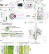
Figure 1:. Muscle EF is distinct from muscle tissue and serum proteome and can serve as potential discovery tool for secreted proteins in muscle tissue.
(A) 1. Scheme of EF in the muscle tissue. SF = Secreted Factor 2. Scheme of muscle EF isolation procedure. (B) Scheme of MS experiment comparing I. EF to muscle tissue and II. EF to serum (C) Principal component analysis of I. EF vs. muscle tissue and II. EF vs. serum (n = 5–6 per compartment). (D) Venn diagram of proteins increased in EF and muscle tissue. Proteins up/down determined by q-value < 0.05. (E) Fishers exact test of secreted proteins in EF and muscle compared to UniProt dataset. (F) Venn diagram of proteins increased in EF and serum. Proteins up/down determined by q-value < 0.05. See also Figure S1 and Table S1 and S2.
Figure 2:
Figure 2:. Acute exercise training remodels the muscle EF proteome and impacts proteins of the coagulation and complement cascades.
(A) Scheme of acute exercise and EF isolation procedure and processing. (B) Western blot of GFP protein expression in muscle EF and tissue of sedentary and exercised mice 2 weeks post GFP adenovirus intramuscular injection. (C) Western blot analysis of common intracellular marker proteins in muscle EF and tissue of sedentary and exercised mice. (D) ELISA of EF IL-6 levels in sedentary and exercised mice (two-tailed unpaired t-test, n = 6). (E) Venn diagram of quantified proteins and significantly changed proteins upon acute exercise regime (significant if q-value < 0.05, n = 5–6 per group). (F) KEGG pathway analysis of significantly downregulated proteins annotated as secreted in exercise. (G) Volcano plot of top 10 upregulated and downregulated proteins annotated as secreted (significant if q-value < 0.05, n = 5–6). (H) Heatmap of top 10 upregulated and downregulated proteins annotated as secreted (q-value < 0.05, n = 5–6). Data are presented as means ± S.E.M. See also Figure S2 and Table S3 and S4.
Figure 3:
Figure 3:. Muscle-specific PGC1α expression remodels EF proteome and reveals elevated protein levels of the neurotrophic factor prosaposin.
(A) Scheme of MCK-PGC1α EF isolation procedure and processing. (B) qRT-PCR of Ppargc1a gene expression normalized to rplp0 in different tissues (two-way ANOVA, n = 4). Gas = Gastrocnemius muscle, BAT = brown adipose tissue. (C) Venn diagram of quantified proteins and significantly changed proteins in MCK-PGC1α vs. ctrl EF (significant if q-value < 0.05, n = 5 per genotype). (D) Scheme of data filtering procedure. (E) Volcano plot of top 10 up- and downregulated proteins annotated as secreted (significant if q-value < 0.05, n = 5). (F) Heatmap of top 10 upregulated and downregulated proteins annotated as secreted (log2 fold changes, n = 5). Data are presented as means ± S.E.M. See also Figure S3 and Table S5.
Figure 4:
Figure 4:. EF proteome analysis identifies many cold-induced, secreted factors in thermogenic adipose tissue, including PSAP.
(A) Scheme of fat depot EF isolation procedure and processing. (B) qRT-PCR of thermogenic gene expression normalized to rplp0 in iWAT after 2 weeks room temperature or cold exposure (unpaired t-test, n = 4–5). (C) Venn diagram of quantified proteins and significantly changed proteins in cold-adapted EF of iWAT vs. eWAT (significant if q-value < 0.05, n = 4, EF pooled from 5 mice per sample). (D) Scheme of data filtering procedure including overlay of upregulated proteins annotated as secreted in iWAT EF vs. upregulated mRNA in TRAP dataset . (E) Volcano plot of top 15 upregulated proteins annotated as secreted in cold-adapted EF of iWAT vs. eWAT (significant if q-value < 0.05, n = 4, EF pooled from 5 mice per sample). (F) Venn diagram of proteins annotated as secreted and significantly changed upon cold exposure in iWAT EF and PGC1α expression in muscle EF (significant if q-value < 0.05). Data are presented as means ± S.E.M. See also Figure S4 and Table S6.
Figure 5:
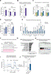
Figure 5:. PSAP expression and secretion is induced by PGC1α and cold adaptation in muscle and fat and PSAP expression is sufficient to boost oxidative metabolism in primary iWAT adipocytes.
(A) qRT-PCR of Psap gene expression normalized to rplp0 in different tissues of MCK-PGC1α mice (two-way ANOVA, n = 4). Gas = gastrocnemius muscle, BAT = brown adipose tissue. (B) PSAP intensity in conditioned medium of PGC1α- and GFP-transduced primary myotubes (unpaired t-test, n = 4). (C) qRT-PCR of Psap gene expression normalized to rplp0 in different fat depots upon thermoneutrality (30 °C), room temperature (22 °C), or cold exposure (4 °C) (one-way ANOVA, n = 4–5). iWAT = inguinal white adipose tissue, BAT = brown adipose tissue, eWAT = epididymal white adipose tissue. (D) qRT-PCR of Psap gene expression in iWAT and BAT upon different times of cold exposure (one-way ANOVA, n = 4–5). iWAT = inguinal white adipose tissue, BAT = brown adipose tissue. (E) qRT-PCR of thermogenic gene expression and brown-fat identity genes normalized to rplp0 in primary iWAT adipocytes transduced with GFP- or PSAP-adenovirus (pAd) (two-way ANOVA, n = 4). (F) Western blot of UCP1 protein expression in primary iWAT adipocytes transduced with GFP- or PSAP-pAd (n = 4). (G) Top upregulated hallmarks in GSEA enrichment analysis of RNAseq data of primary iWAT adipocytes transduced with GFP- or PSAP-pAd. (H) Enrichment plot of top upregulated hallmark oxidative phosphorylation in GSEA enrichment analysis of RNAseq data from primary iWAT adipocytes transduced with GFP- or PSAP-pAd (q-value < 0.001, n = 4). (I) Oxygen consumption rate (OCR) of iWAT adipocytes transduced with GFP- or PSAP-pAd (n = 10). NE = norepinephrine, Oligo = oligomycin, FCCP = carbonilcyanide p-triflouromethoxyphenylhydrazone, Rot = rotenone, Anti A = antimycin A (J) Basal OCR of iWAT adipocytes transduced with PSAP or GFP adenovirus (two-tailed unpaired t-test, n = 10). Data are presented as means ± S.E.M. See also Table S7.

References

    1. Severinsen MCK, and Pedersen BK (2020). Muscle-Organ Crosstalk: The Emerging Roles of Myokines. Endocr Rev 41. 10.1210/endrev/bnaa016. - DOI - PMC - PubMed
    1. de Oliveira Dos Santos AR, de Oliveira Zanuso B, Miola VFB, Barbalho SM, Santos Bueno PC, Flato UAP, Detregiachi CRP, Buchaim DV, Buchaim RL, Tofano RJ, et al. (2021). Adipokines, Myokines, and Hepatokines: Crosstalk and Metabolic Repercussions. Int J Mol Sci 22. 10.3390/ijms22052639. - DOI - PMC - PubMed
    1. Leuchtmann AB, Adak V, Dilbaz S, and Handschin C (2021). The Role of the Skeletal Muscle Secretome in Mediating Endurance and Resistance Training Adaptations. Front Physiol 12, 709807. 10.3389/fphys.2021.709807. - DOI - PMC - PubMed
    1. Villarroya J, Cereijo R, Gavalda-Navarro A, Peyrou M, Giralt M, and Villarroya F (2019). New insights into the secretory functions of brown adipose tissue. J Endocrinol 243, R19–R27. 10.1530/JOE-19-0295. - DOI - PubMed
    1. Murphy RM, Watt MJ, and Febbraio MA (2020). Metabolic communication during exercise. Nat Metab 2, 805–816. 10.1038/s42255-020-0258-x. - DOI - PubMed

Publication types