. 2023 Mar 1:462:116381.
doi: 10.1016/j.taap.2023.116381. Epub 2023 Jan 19.

Scavenger receptor BI attenuates oxidized phospholipid-induced pulmonary inflammation

Affiliations

Scavenger receptor BI attenuates oxidized phospholipid-induced pulmonary inflammation

Katelyn Dunigan-Russell et al. Toxicol Appl Pharmacol. .

Abstract

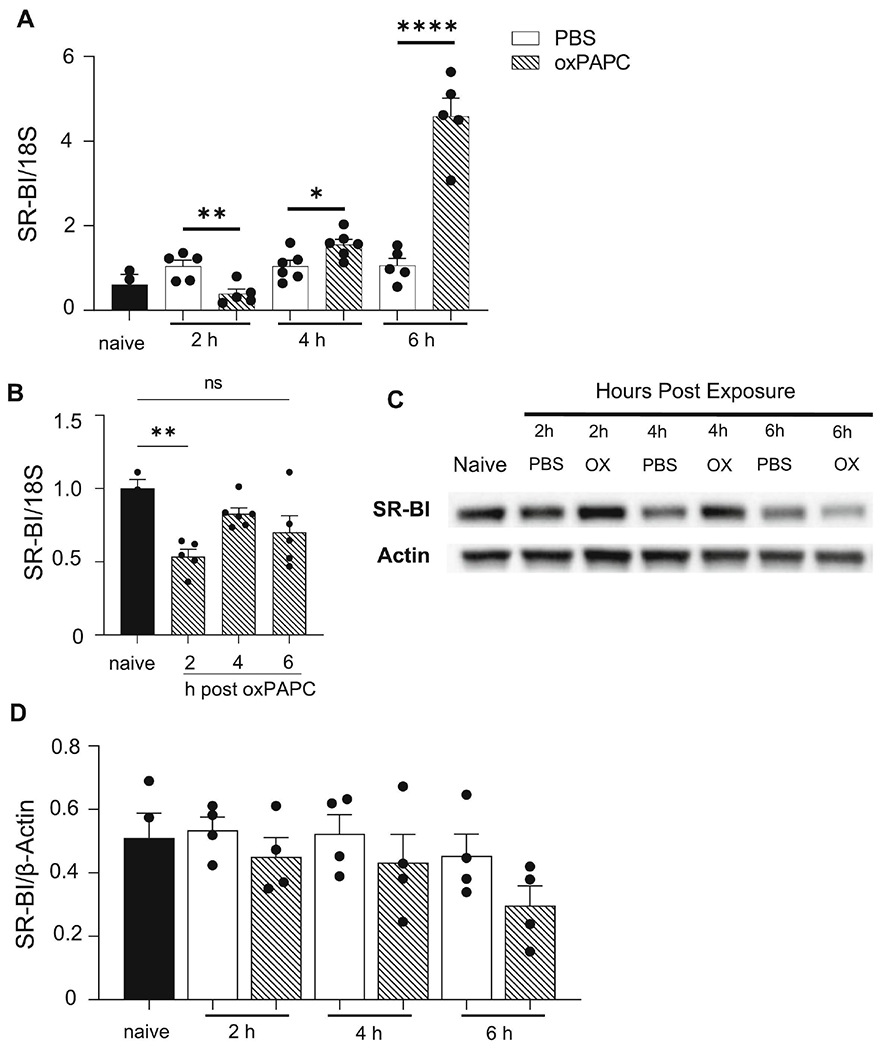
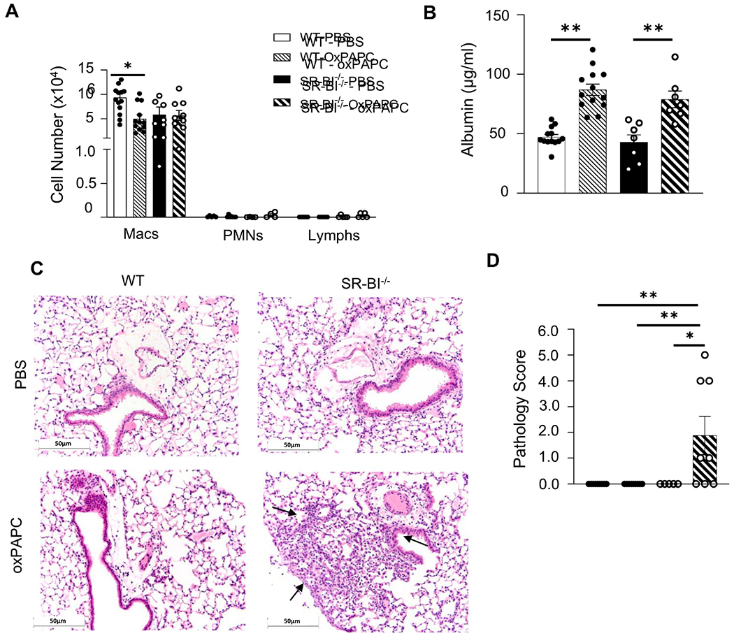
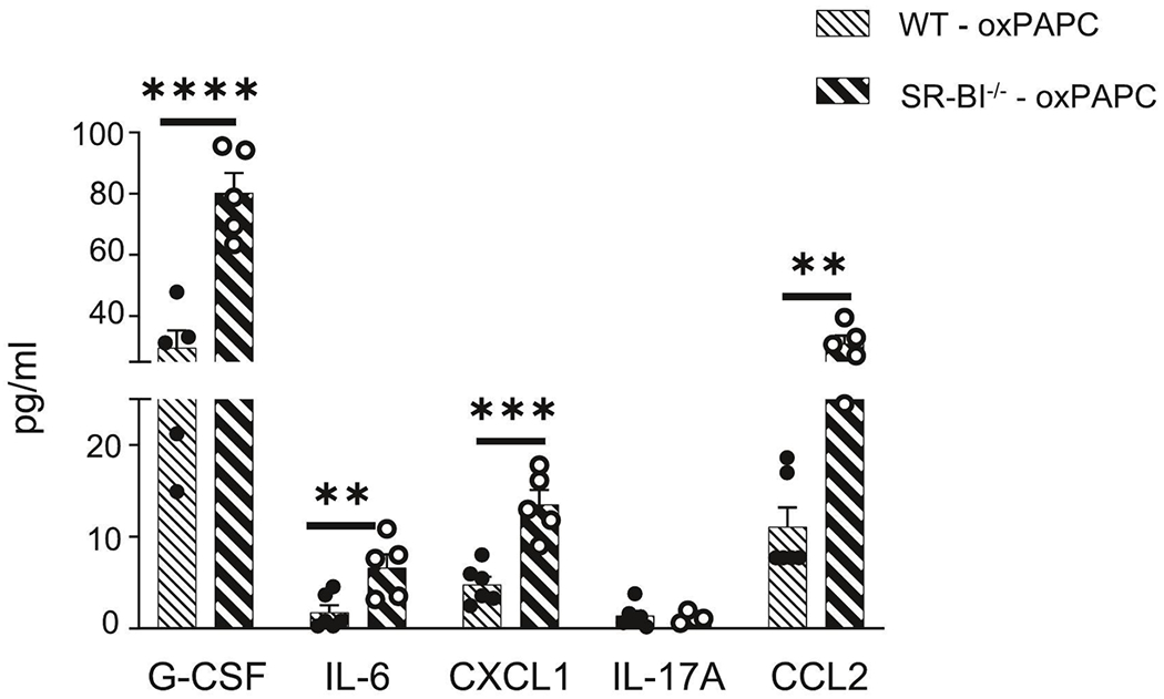
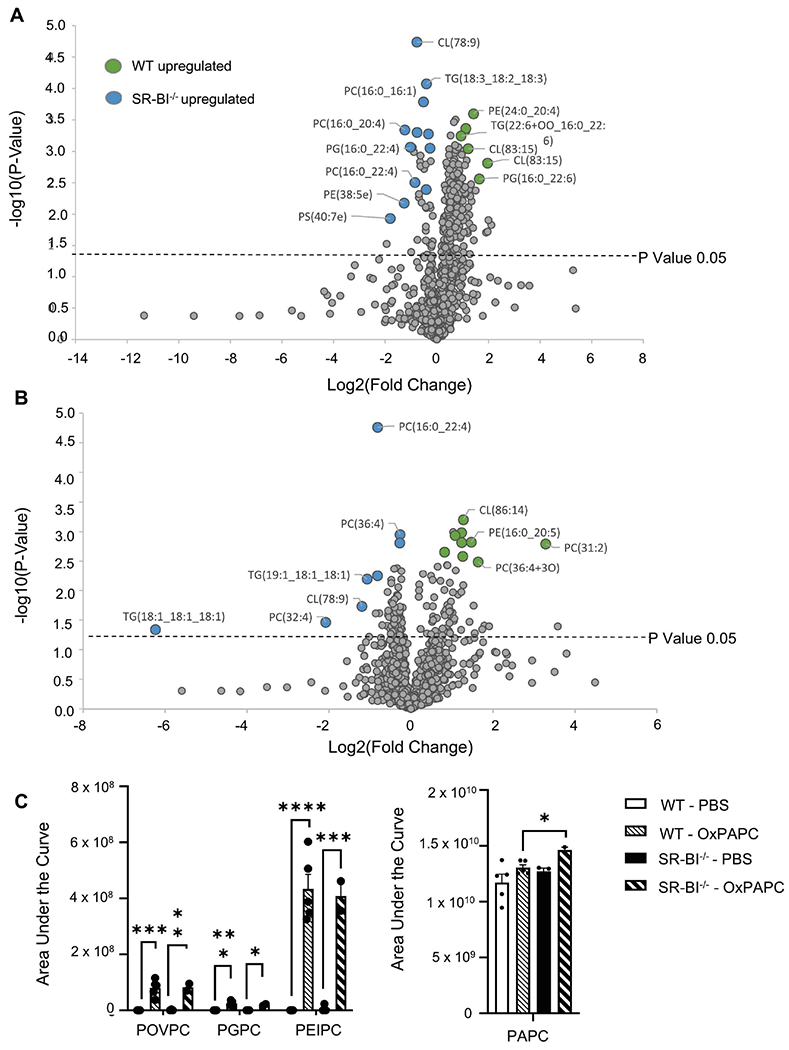
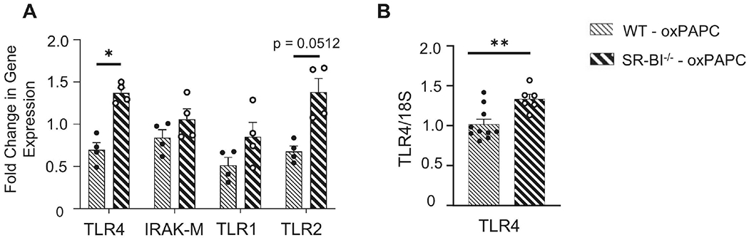
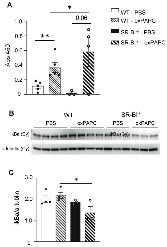
Damage associated molecular patterns (DAMPs) are molecules released from dead/dying cells following toxicant and/or environmental exposures that activate the immune response through binding of pattern recognition receptors (PRRs). Excessive production of DAMPs or failed clearance leads to chronic inflammation and delayed inflammation resolution. One category of DAMPs are oxidized phospholipids (oxPLs) produced upon exposure to high levels of oxidative stress, such as following ozone (O3) induced inflammation. OxPLs are bound by multiple classes of PRRs that include scavenger receptors (SRs) such as SR class B-1 (SR-BI) and toll-like receptors (TLRs). Interactions between oxPLs and PRRs appear to regulate inflammation; however, the role of SR-BI in oxPL-induced lung inflammation has not been defined. Therefore, we hypothesize that SR-BI is critical in protecting the lung from oxPL-induced pulmonary inflammation/injury. To test this hypothesis, C57BL/6J (WT) female mice were dosed with oxidized 1-palmitoyl-2-arachidonoyl-sn-glycero-3-phosphatidylcholine (oxPAPC) by oropharyngeal aspiration which increased pulmonary SR-BI expression. Following oxPAPC exposure, SR-BI deficient (SR-BI-/-) mice exhibited increased lung pathology and inflammatory cytokine/chemokine production. Lipidomic analysis revealed that SR-BI-/- mice had an altered pulmonary lipidome prior to and following oxPAPC exposure, which correlated with increased oxidized phosphatidylcholines (PCs). Finally, we characterized TLR4-mediated activation of NF-κB following oxPAPC exposure and discovered that SR-BI-/- mice had increased TLR4 mRNA expression in lung tissue and macrophages, increased nuclear p65, and decreased cytoplasmic IκBα. Overall, we conclude that SR-BI is required for limiting oxPAPC-induced lung pathology by maintaining lipid homeostasis, reducing oxidized PCs, and attenuating TLR4-NF-κB activation, thereby preventing excessive and persistent inflammation.

Keywords: DAMP; Inflammation; Lung; SR-BI; TLR4.


Conflict of interest statement

Declaration of Competing Interest The authors declare that they have no known competing financial interests or personal relationships that could have appeared to influence the work reported in this paper.

Figures

Figure 1.
Figure 1.. SR-BI gene expression is increased in the lungs after oxPAPC exposure.
Female WT mice were unexposed (naïve), exposed to PBS, or oxPAPC via oropharyngeal (o.p.) aspiration and then euthanized 2, 4, or 6 h post- exposure. Lung tissue was collected to assess SR-BI (A) mRNA gene expression by qPCR, and (B) protein via western blot, which was quantified by densitometry and normalized to β-actin. *p<0.05, **p<0.01, ****p<0.0001; n=3-5 per group.
Figure 2.
Figure 2.. SR-BI deficiency increases susceptibility to oxPAPC-induced lung injury.
Female WT or SR-BI−/− mice were exposed to PBS or oxPAPC via oropharyngeal (o.p.) aspiration and euthanized 4 h after exposure. Bronchoalveolar lavage (BAL) was collected for (A) cell differentials, and (B) albumin analysis. Lungs were then fixed with 10% neutral-buffered formalin and stained with (C) H&E and were (D) graded by a blinded board-certified veterinary pathologist. Arrows to indicate areas of pulmonary injury. Representative images are at 20x magnification. **p<0.01, ***p<0.001, ****p<0.0001; n=5-14 per group.
Figure 3.
Figure 3.. Loss of SR-BI increases cyto/chemokine production in the airspace after oxPAPC exposure.
Female WT or SR-BI−/− mice were exposed to oxPAPC via oropharyngeal (o.p.) aspiration and euthanized 4 h after exposure. Bronchoalveolar lavage (BAL) was collected for multiplex analysis of G-CSF, IL-6, CXCL1, IL-17A, and CCL2. *p<0.05, **p<0.01, ***p<0.001; n=5-13 per group.
Figure 4.
Figure 4.. SR-BI pharmacological inhibition increases susceptibility to oxPAPC-induced lung injury.
Female WT mice were exposed to PBS or BLT-2 (SR-BI inhibitor) 1 h prior to PBS or oxPAPC exposure. Then, 4 h following exposure, mice were euthanized and bronchoalveolar lavage (BAL) was collected to measure (A) cell differentials, (B) protein, and (C) cytokines/chemokines via multiplex analysis. Lungs were also fixed with 10% neutral-buffered formalin and stained for (D) H&E and were (E) graded by a blinded board-certified veterinary pathologist. Arrows to indicate areas of pulmonary injury. Representative images are at 20x magnification. *p<0.05, **p<0.01; n=3-15 per group.
Figure 5.
Figure 5.. OxPLs are increased in the lungs of SR-BI−/− mice.
Female WT or SR-BI−/− mice were exposed to PBS or oxPAPC via oropharyngeal (o.p.) aspiration and were euthanized 4 h after exposure. Bronchoalveolar lavage (BAL) was collected for LC-MS/MS analysis. (A) Relative phospholipid concentrations were compared between WT and SR-BI−/− mice after PBS exposure and (B) after OxPAPC exposure. Increased fold change values for WT animals are shaded green while increased fold change values in SR-BI−/− mice are shaded blue and (C) the area under the curve quantitation of relative concentrations of oxidized lipid species POVPC, PGPC, PEIPC, and PAPC normalized to a deuterated PC internal standard (PC-IS). *p<0.05, **p<0.01, ***p<0.001, ****p<0.0001; n=3-6 per group.
Figure 6.
Figure 6.. TLR4 expression is upregulated in SR-BI−/− mice after oxPAPC exposure.
Female WT or SR-BI−/− mice were exposed to oxPAPC via oropharyngeal (o.p.) aspiration and euthanized 4 h after exposure. (A) Lung tissue was collected to analyze mRNA expression TLR4, IRAK-M, TLR1, and TLR2 by qPCR. (B) Macrophages were isolated from bronchoalveolar lavage (BAL), lysed, and mRNA expression of TLR4 was analyzed by qPCR. *p<0.05; n=4-10 per group.
Figure 7.
Figure 7.. NF-κB activation is upregulated in SR-BI−/− mice after oxPAPC exposure.
Female WT or SR-BI−/− mice were exposed to PBS or oxPAPC via oropharyngeal (o.p.) aspiration and euthanized 2h after exposure. (A) Lung tissue was collected and nuclear-cytoplasmic fractionation was conducted to measure p65 in the nuclear fraction via p65 ELISA. (B) Female WT or SR-BI−/− mice were exposed to PBS or oxPAPC via oropharyngeal (o.p.) aspiration and euthanized 4h after exposure. Lung tissue was collected and nuclear-cytoplasmic fractionation was conducted to measure cytoplasmic (cy) IκBα via western blot. (C) Densitometry of the cytoplasmic (cy) IκBα relative to α-tubulin. *p<0.05, **p<0.01; n=3-5 per group.

References

    1. Gong T, Liu L, Jiang W, Zhou R. DAMP-sensing receptors in sterile inflammation and inflammatory diseases. Nat Rev Immunol. 2020;20(2):95–112. - PubMed
    1. Stamenkovic A, Pierce GN, Ravandi A. Oxidized lipids: not just another brick in the wall (1). Can J Physiol Pharmacol. 2019;97(6):473–85. - PubMed
    1. Minagawa S, Yoshida M, Araya J, Hara H, Imai H, Kuwano K. Regulated Necrosis in Pulmonary Disease. A Focus on Necroptosis and Ferroptosis. Am J Respir Cell Mol Biol. 2020;62(5):554–62. - PubMed
    1. Ito K, Eguchi Y, Imagawa Y, Akai S, Mochizuki H, Tsujimoto Y. MPP+ induces necrostatin-1- and ferrostatin-1-sensitive necrotic death of neuronal SH-SY5Y cells. Cell Death Discov. 2017;3:17013. - PMC - PubMed
    1. Kuwano K, Kunitake R, Kawasaki M, Nomoto Y, Hagimoto N, Nakanishi Y, et al. P21Waf1/Cip1/Sdi1 and p53 expression in association with DNA strand breaks in idiopathic pulmonary fibrosis. Am J Respir Crit Care Med. 1996;154(2 Pt 1):477–83. - PubMed

Publication types