

Protracted development of motor cortex constrains rich interpretations of infant cognition
- PMID: 36681607
- PMCID: PMC9957955
- DOI: 10.1016/j.tics.2022.12.014
Protracted development of motor cortex constrains rich interpretations of infant cognition
Abstract
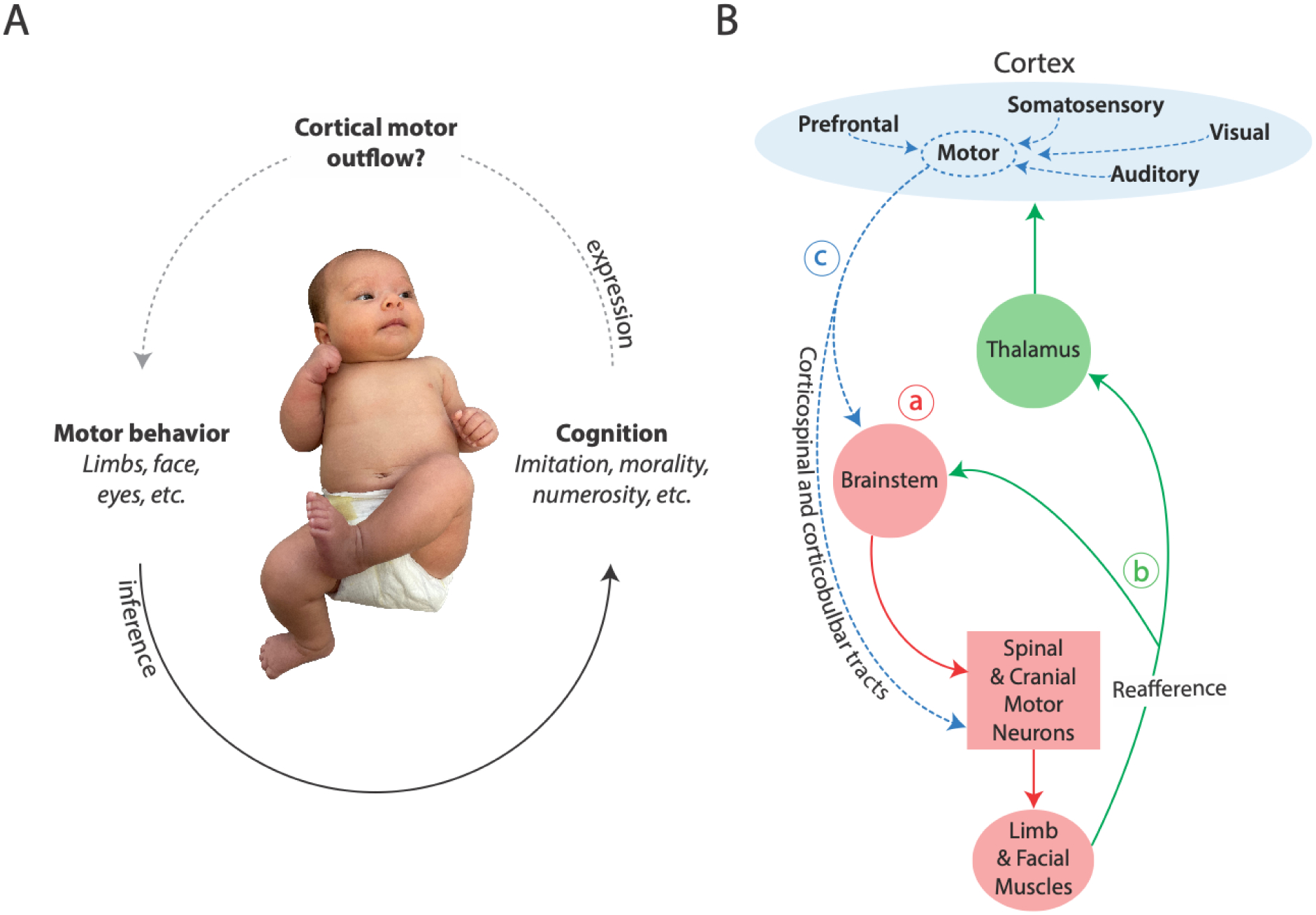
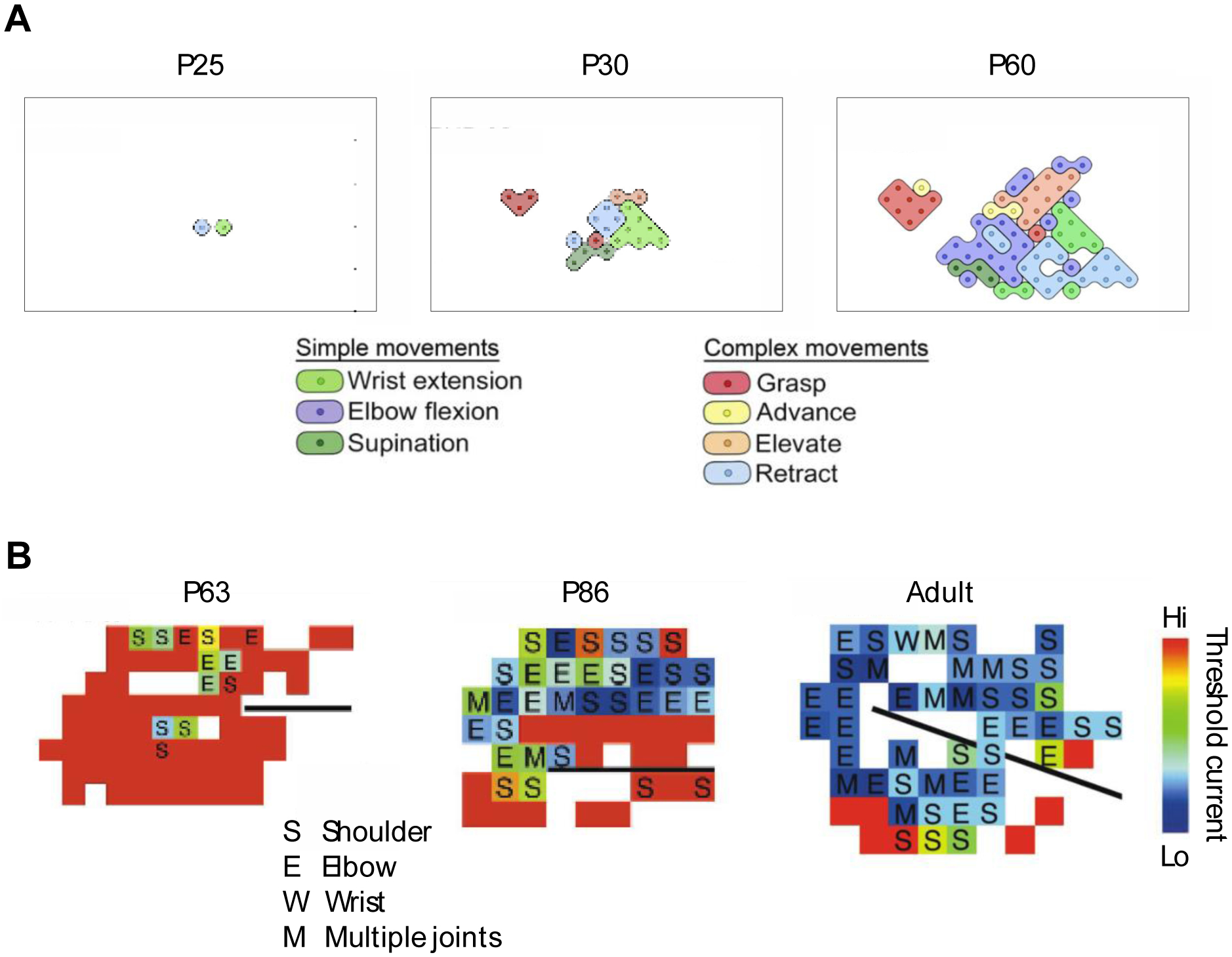
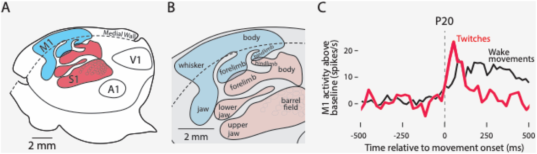
Cognition in preverbal human infants must be inferred from overt motor behaviors such as gaze shifts, head turns, or reaching for objects. However, infant mammals - including human infants - show protracted postnatal development of cortical motor outflow. Cortical control of eye, face, head, and limb movements is absent at birth and slowly emerges over the first postnatal year and beyond. Accordingly, the neonatal cortex in humans cannot generate the motor behaviors routinely used to support inferences about infants' cognitive abilities, and thus claims of developmental continuity between infant and adult cognition are suspect. Recognition of the protracted development of motor cortex should temper rich interpretations of infant cognition and motivate more serious consideration of the role of subcortical mechanisms in early cognitive development.
Keywords: core knowledge; eye movements; infant cognition; motor cortex; motor maps; visual system.
Copyright © 2023 Elsevier Ltd. All rights reserved.
Conflict of interest statement
Declaration of interests The authors have no interests to declare.
Figures
Comment in
-
Infant action and cognition: what's at stake?Trends Cogn Sci. 2023 Aug;27(8):696-698. doi: 10.1016/j.tics.2023.05.008. Epub 2023 Jun 13. Trends Cogn Sci. 2023. PMID: 37321923 Free PMC article. No abstract available.
-
Multiple pathways to developmental continuity in infant cognition.Trends Cogn Sci. 2023 Aug;27(8):692-693. doi: 10.1016/j.tics.2023.04.007. Epub 2023 Jun 13. Trends Cogn Sci. 2023. PMID: 37321924 No abstract available.
Similar articles
-
The developmental origins of naïve psychology in infancy.Adv Child Dev Behav. 2009;37:55-104. doi: 10.1016/s0065-2407(09)03702-1. Adv Child Dev Behav. 2009. PMID: 19673160 Review.
-
Morphological features of the neonatal brain support development of subsequent cognitive, language, and motor abilities.Hum Brain Mapp. 2014 Sep;35(9):4459-74. doi: 10.1002/hbm.22487. Epub 2014 Feb 25. Hum Brain Mapp. 2014. PMID: 24615961 Free PMC article.
-
Visual fixation in human newborns correlates with extensive white matter networks and predicts long-term neurocognitive development.J Neurosci. 2015 Mar 25;35(12):4824-9. doi: 10.1523/JNEUROSCI.5162-14.2015. J Neurosci. 2015. PMID: 25810513 Free PMC article.
-
Early exploration of one's own body, exploration of objects, and motor, language, and cognitive development relate dynamically across the first two years of life.Dev Psychol. 2022 Feb;58(2):222-235. doi: 10.1037/dev0001289. Epub 2022 Jan 6. Dev Psychol. 2022. PMID: 34990201 Free PMC article.
-
Close interrelation of motor development and cognitive development and of the cerebellum and prefrontal cortex.Child Dev. 2000 Jan-Feb;71(1):44-56. doi: 10.1111/1467-8624.00117. Child Dev. 2000. PMID: 10836557 Review.
Cited by
-
Perceptual and conceptual novelty independently guide infant looking behaviour: a systematic review and meta-analysis.Nat Hum Behav. 2024 Dec;8(12):2342-2356. doi: 10.1038/s41562-024-01965-x. Epub 2024 Oct 14. Nat Hum Behav. 2024. PMID: 39402259
-
A hierarchical model of early brain functional network development.Trends Cogn Sci. 2025 May 6:S1364-6613(25)00080-4. doi: 10.1016/j.tics.2025.04.001. Online ahead of print. Trends Cogn Sci. 2025. PMID: 40335413 Review.
-
What we don't know about what babies know: Reconsidering psychophysics, exploration, and infant behavior.Behav Brain Sci. 2024 Jun 27;47:e121. doi: 10.1017/S0140525X23003217. Behav Brain Sci. 2024. PMID: 38934452 Free PMC article.
-
Genome-wide association meta-analysis of age at onset of walking in over 70,000 infants of European ancestry.Nat Hum Behav. 2025 Jul;9(7):1470-1487. doi: 10.1038/s41562-025-02145-1. Epub 2025 May 7. Nat Hum Behav. 2025. PMID: 40335706 Free PMC article.
-
Consciousness in the cradle: on the emergence of infant experience.Trends Cogn Sci. 2023 Dec;27(12):1135-1149. doi: 10.1016/j.tics.2023.08.018. Epub 2023 Oct 12. Trends Cogn Sci. 2023. PMID: 37838614 Free PMC article. Review.
References
-
- Spelke ES and Kinzler KD (2007) Core knowledge. Dev Sci 10, 89–96 - PubMed
-
- Hamlin JK (2013) Moral judgment and action in preverbal infants and toddlers: Evidence for an innate moral core. Curr Dir Psychol Sci 22, 186–193
-
- Haith M (1998) Who put the cog in infant cognition? Is rich interpretation too costly. Infant Behav Dev 21, 167–179
Publication types
MeSH terms
Grants and funding
LinkOut - more resources
Full Text Sources
