. 2023 Jan 21;9(1):21.
doi: 10.1038/s41420-023-01330-3.

DNA damage triggers squamous metaplasia in human lung and mammary cells via mitotic checkpoints

Affiliations

DNA damage triggers squamous metaplasia in human lung and mammary cells via mitotic checkpoints

Lucía San Juan et al. Cell Death Discov. .

Abstract

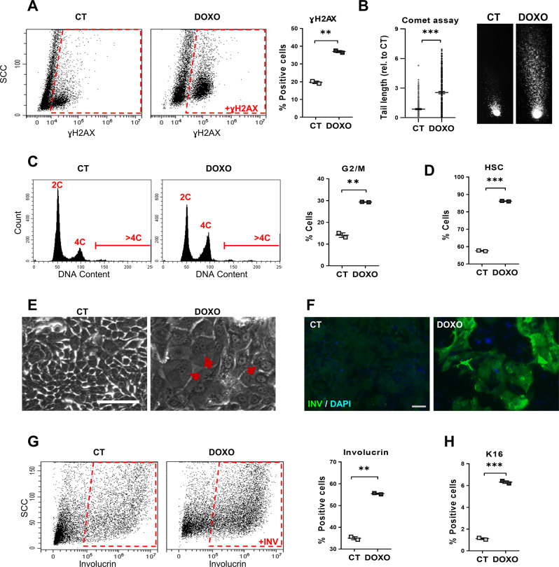
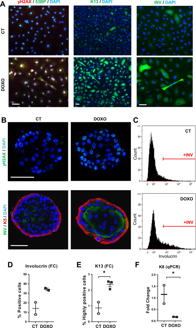
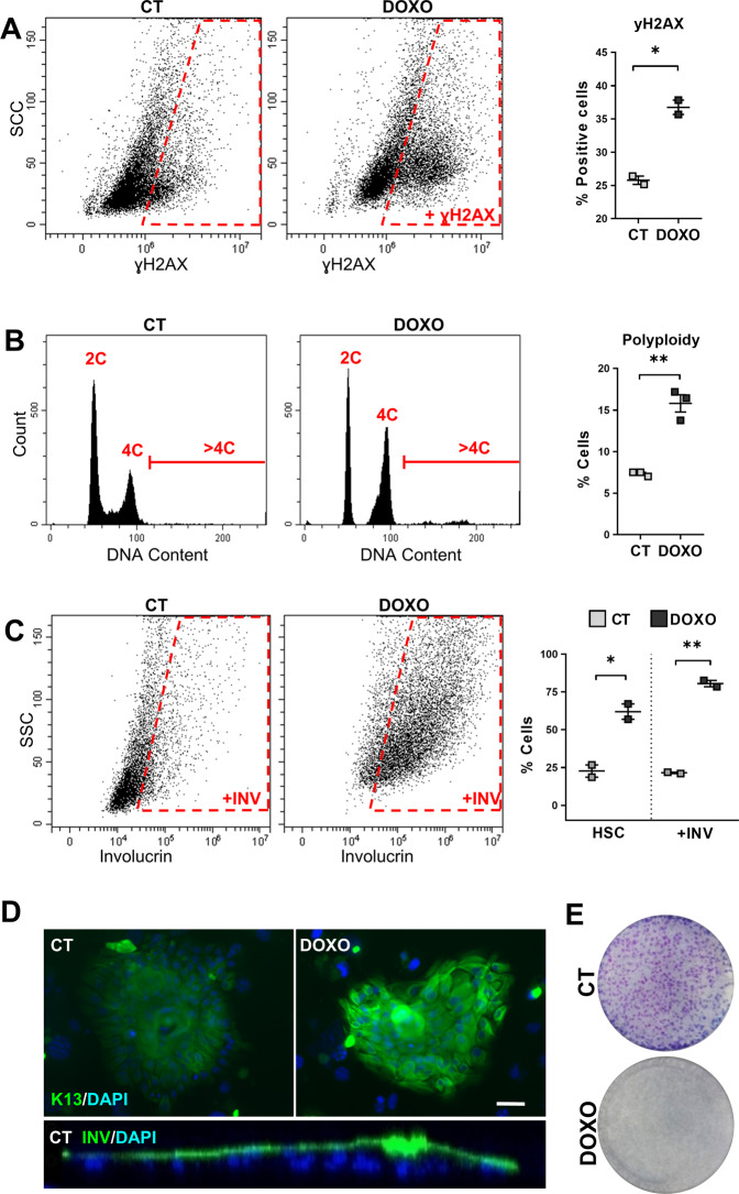
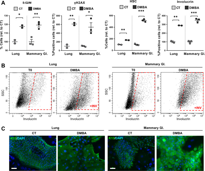
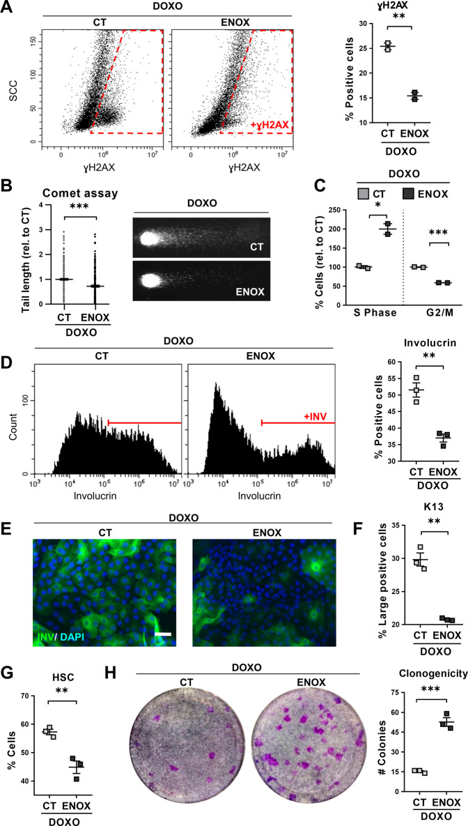
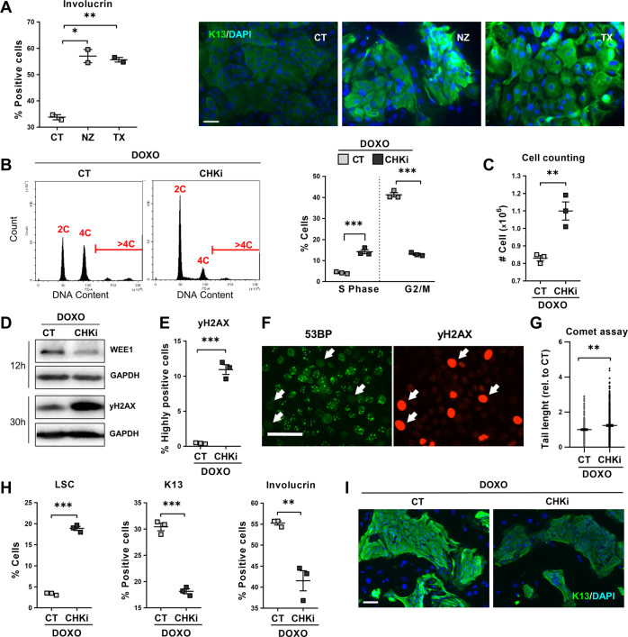
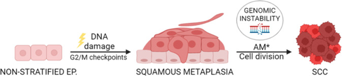
Epithelial transdifferentiation is frequent in tissue hyperplasia and contributes to disease in various degrees. Squamous metaplasia (SQM) precedes epidermoid lung cancer, an aggressive and frequent malignancy, but it is rare in the epithelium of the mammary gland. The mechanisms leading to SQM in the lung have been very poorly investigated. We have studied this issue on human freshly isolated cells and organoids. Here we show that human lung or mammary cells strikingly undergo SQM with polyploidisation when they are exposed to genotoxic or mitotic drugs, such as Doxorubicin or the cigarette carcinogen DMBA, Nocodazole, Taxol or inhibitors of Aurora-B kinase or Polo-like kinase. To note, the epidermoid response was attenuated when DNA repair was enhanced by Enoxacin or when mitotic checkpoints where abrogated by inhibition of Chk1 and Chk2. The results show that DNA damage has the potential to drive SQM via mitotic checkpoints, thus providing novel molecular candidate targets to tackle lung SCC. Our findings might also explain why SCC is frequent in the lung, but not in the mammary gland and why chemotherapy often causes complicating skin toxicity.


Conflict of interest statement

HC is an inventor of patents related to organoid research, His full disclosure: www.uu.nl/staff/JCClevers/Additional. Otherwise, the authors declare no competing interests.

Figures

Fig. 1
Fig. 1. Doxorubicin induces squamous metaplasia in human lung epithelial cells.
Human primary lung epithelial cells were treated with the dimethyl sulfoxide vehicle (CT) or with 0.5 μM Doxorubicin (DOXO) for 24 h (A, C) or 48 h (B, DH). A Representative flow-cytometry analysis for the DNA damage marker γH2AX (+γH2AX, positive cells). Quantitation in the right histogram. B DNA fragmentation as analysed by comet assays, measured by tail length relative to CT (n = 245–247). Photographs show representative images of nuclei in CT or DOXO-treated cells as indicated. C Representative flow-cytometry analyses of DNA content of cells (2C, 4C, and >4C indicate diploid, mitotic/tetraploid and polyploid cells, respectively). Plot on the right shows percent of CT or DOXO cells in the G2/M phase of the cell cycle. D Percent of CT or DOXO-treated cells with high light scatter (HSC) typical of squamous differentiation, as analysed by flow cytometry. E Representative phase contrast images of cells treated for 48 h as indicated. Red arrows point at polyploid cells with several or large nuclei. Scale bar, 50 μm. F Immunofluorescence for the squamous differentiation marker involucrin (green). Blue is nuclear DNA by DAPI. Scale bar, 100 μm. G Representative flow cytometry analysis for involucrin (+INV, positive cells). The percent of involucrin positive CT or DOXO-treated cells is shown on the right. H Percent of squamous marker keratin K16 positive cells, as analysed by flow cytometry. Flow cytometry analysis gates were stablished according to negative isotype antibody control. Data are mean ± SEM of duplicate samples, representative of 2–3 independent experiment from two different human individuals with similar results. ***p ≤ 0.001, **p ≤ 0.01.
Fig. 2
Fig. 2. Doxorubicin induces squamous differentiation in human lung epithelial cells in a serum-free lung-adapted medium and in organoid reconstructions.
A Human primary lung epithelial cells isolated and cultured in lung-adapted medium were treated for 48 h with the dimethyl sulfoxide vehicle (CT) or with 0.5 μM Doxorubicin (DOXO), as indicated. Immunofluorescence for 53BP (green) and γH2AX (red), for K13 (green), or for involucrin (green), as indicated. Blue is nuclear DNA by DAPI; scale bar, 50 μm. Representative of two independent experiments with similar results. BF Airway lung organoids (AOs) in organoid expansion medium were treated with dimethyl sulfoxide (CT) or with 0.25 μM Doxorubicin (DOXO) for 24 h (C, D, F) or 48 h (B, E), as indicated. B Immunofluorescence confocal images of AOs treated as indicated and stained for γH2AX (top; green) or for involucrin (bottom; green) and K5 (red). Scale bar, 50 μm. Blue is nuclear DNA by DAPI. C Representative flow cytometry analyses for the differentiation marker involucrin (+INV, positive cells). D Percent of involucrin positive cells by flow cytometry. E Percent of keratin K13 positive cells by flow cytometry. F mRNA fold change of keratin K8 by qPCR. Positive cells by flow cytometry were gated according to negative isotype antibody control. Data are mean ± SEM of two or three replicate samples of two independent experiments from two different human individual. *p ≤ 0.05, p for +INV = 0.09.
Fig. 3
Fig. 3. Doxorubicin induces squamous metaplasia in human mammary gland epithelial cells.
Human primary mammary gland epithelial cells were treated with dimethyl sulfoxide (CT) or with 0.5 μM Doxorubicin (DOXO) for 48 h, as indicated. A Representative flow cytometry analysis for the DNA damage marker γH2AX (+γH2AX, positive cells). The percent of +γH2AX is shown on the right. B Representative flow cytometry analyses of DNA content (2C, 4C, and >4C). The percent of polyploid cells (>4C) is shown on the right. C Representative flow-cytometry analysis for the differentiation marker involucrin (+INV, positive cells). The percent of cells with high light scatter (HSC), or +INV is shown on the right, as indicated. D Top: representative mammary gland colonies after immunofluorescence for keratin K13 (green), treated as indicated. Blue is nuclear DNA by DAPI. Scale bar, 50 μm. Bottom: Orthogonal view profile of a CT mammary gland colony after immunofluorescence for involucrin (INV). E Clonogenic capacity of cells plated after 48 h treatment as indicated and drug-released. Positive cells by flow cytometry were gated according to negative isotype antibody control. Data are mean ± SEM of duplicate or triplicate samples, representative of two independent experiment from two different human individuals with similar results. **p ≤ 0.01, *p ≤ 0.05.
Fig. 4
Fig. 4. Lung and mammary gland epithelial cells undergo squamous metaplasia upon tobacco carcinogen DMBA.
Human primary lung or mammary gland (Mammary Gl.) epithelial cells were treated with dimethyl sulfoxide (CT) or with 1 μg/ml DMBA for 24 h (A, S-G2/M, and yH2AX) or 72 h (A, HSC and involucrin, B, C). A Flow cytometry quantitations for the percent of, from left to right: cells in the S-G2/M phase of the cell cycle, γH2AX positive cells, cells with high light scatter (HSC) typical of squamous differentiation, involucrin positive cells, all relative to CT. B Representative flow cytometry analyses for the differentiation marker involucrin. +INV, positive cells; T0 = untreated cells. C Representative immunofluorescence for keratin K13 (green) upon the indicated treatments. Blue is nuclear DNA by DAPI. Scale bar, 50 μm. Positive cells by flow cytometry were gated according to negative isotype antibody control. Data are mean ± SEM of two or three replicate samples, representative of 2–3 independent experiment from two different human individuals with similar results. ***p ≤ 0.001, **p ≤ 0.01, *p ≤ 0.05.
Fig. 5
Fig. 5. Enhancement of DNA repair by Enoxacin suppresses Doxorubicin-induced squamous metaplasia in lung epithelial cells.
Human primary lung epithelial cells were treated with Doxorubicin (DOXO) and with the dimethyl sulfoxide vehicle (CT) or with 200 μM Enoxacin (ENOX). A Representative flow-cytometry analyses for the DNA damage marker γH2AX of cells treated for 24 h, as indicated (γH2AX+, positive cells). Scattered plot (right) shows percent of γH2AX positive cells. B DNA fragmentation as analysed by comet assays after 24 h treatment as indicated and measured by tail length relative to CT (n = 237–312). Photographs show representative images of nuclei in CT or ENOX-treated cells, as indicated. C Percent of cells in the S or G2/M phases of the cell cycle relative to CT, analysed by flow cytometry after 24 h with the indicated treatments. D Representative flow cytometry analyses for involucrin (+INV, positive cells) after 48 h with the indicated treatments. Positive cells were gated according to negative isotype antibody control. The percent of involucrin positive CT or ENOX-treated cells is shown on the right. E Immunofluorescence for involucrin (green) of cells treated for 48 h, as indicated. Blue is nuclear DNA by DAPI. Scale bar, 50 μm. F Percent of large keratin K13 positive cells analysed by flow cytometry after 24 h. G Percent of cells with high light scatter (HSC), analysed by flow cytometry after 24 h. H Clonogenic capacity of cells drug-released and plated after 48 h with the indicated treatments. The number of CT or ENOX colonies is shown on the right plot. Positive cells by flow cytometry were gated according to negative isotype antibody control. Data are mean ± SEM of 2–3 replicate samples, representative of 2–3 independent experiment from two different human individuals with similar results. ***p ≤ 0.001, **p ≤ 0.01, *p ≤ 0.05.
Fig. 6
Fig. 6. The G2/M checkpoints drive squamous metaplasia in response to DNA damage.
A Human primary lung epithelial cells were treated with the dimethyl sulfoxide vehicle (CT), 20 μM Nocodazole (NZ, microtubule polimerisation inhibitor) or 200 μM Paclitaxel (TX, microtubule depolimerisation inhibitor). Left: Percent of CT, NZ, or TX involucrin positive cells, analysed by flow cytometry. Right: Immunofluorescence for involucrin (green). Blue is nuclear DNA by DAPI. Scale bar, 50 μm. BI Human primary lung epithelial cells were treated with 0.07 μM Doxorubicin (DOXO) and with the dimethyl sulfoxide vehicle (CT) or with 0.5 μM AZD7762 (CHKi, Chk1/Chk2 inhibitor). B Representative flow-cytometry analyses of DNA content after the 24 h treatments as indicated. Plot on the right shows percent of CT or CHKi-treated cells in the S or G2/M phase of the cell cycle, as indicated. C Cell counting of recovered CT and CHKi-treated cells after 48 h treatment. D DNA damage marker γH2AX and Wee1 by Western blotting in cells treated for 12 or 30 h, as indicated. Glyceraldehyde 3-phosphate dehydrogenase (GAPDH) as loading control. E Percent of highly γH2AX positive cells, analysed by flow cytometry after 24 h. F Double immunofluorescence for 53BP (green) and γH2AX (red) in CHKi-treated cells at 24 h. Scale bar, 50 μm. White arrows point at strongly positive γH2AX cells lacking 53BP foci (typical of active DNA repair). G DNA fragmentation as analysed by comet assays after 24 h with the indicated treatments, measured by tail length relative to CT (n = 196–298). H Percent of low light scatter (LSC, left) cells, keratin K13 positive cells (centre) or involucrin positive cells (right), as analysed by flow cytometry (K13, 24 h-treatment; LSC and involucrin, 48 h-treatment). I Immunofluorescence of CT and CHKi-treated cells for keratin K13 (green) after 24 h treatments. Blue is nuclear DNA by DAPI. Scale bar, 100 μm. Positive cells by flow cytometry were gated according to negative isotype antibody control. Data are mean ± SEM of duplicate or triplicate samples, representative of 2–3 independent experiment from two different human individuals with similar results. ***p ≤ 0.001, **p ≤ 0.01, *p ≤ 0.05.
Fig. 7
Fig. 7. Model for the role of DNA-damage and mitotic checkpoints in the development of squamous metaplasia and subsequent squamous cancer in non-stratified epithelia.
Chronic or acute exposure of non-squamous epithelia to genotoxic agents would drive G2/M arrest via the DNA damage response. In turn, G2/M checkpoints would drive benign hyperplastic squamous differentiation (squamous metaplasia). Unrepaired cells would undergo terminal post-mitotic squamous differentiation. In the event of additional mutations (AM) hitting the G2/M checkpoints, cells overcoming the cell division block would proliferate bearing genomic instability, eventually resulting in cancer. Ep: epithelium. Created with BioRender.com.

Similar articles

Cited by

References

    1. Sung H, Ferlay J, Siegel RL, Laversanne M, Soerjomataram I, Jemal A, et al. Global Cancer Statistics 2020: GLOBOCAN estimates of incidence and mortality worldwide for 36 cancers in 185 countries. CA Cancer J Clin. 2021;71:209–49. doi: 10.3322/caac.21660. - DOI - PubMed
    1. Boni R, Schuster C, Nehrhoff B, Burg G. Epidemiology of skin cancer. Neuro Endocrinol Lett. 2002;23(Suppl 2):48–51. - PubMed
    1. Ferlay J, Colombet M, Soerjomataram I, Mathers C, Parkin DM, Pineros M, et al. Estimating the global cancer incidence and mortality in 2018: GLOBOCAN sources and methods. Int J Cancer. 2019;144:1941–53. doi: 10.1002/ijc.31937. - DOI - PubMed
    1. World Cancer Research Fund International, 2022. http://www.wcrf.org.
    1. Goodman AM, Kato S, Chattopadhyay R, Okamura R, Saunders IM, Montesion M, et al. Phenotypic and genomic determinants of immunotherapy response associated with squamousness. Cancer Immunol Res. 2019;7:866–73. doi: 10.1158/2326-6066.CIR-18-0716. - DOI - PMC - PubMed