

Immunological and metabolic characteristics of the Omicron variants infection
- PMID: 36681668
- PMCID: PMC9860238
- DOI: 10.1038/s41392-022-01265-8
Immunological and metabolic characteristics of the Omicron variants infection
Abstract
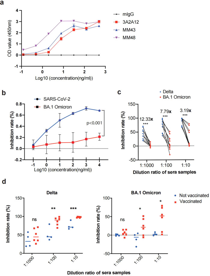
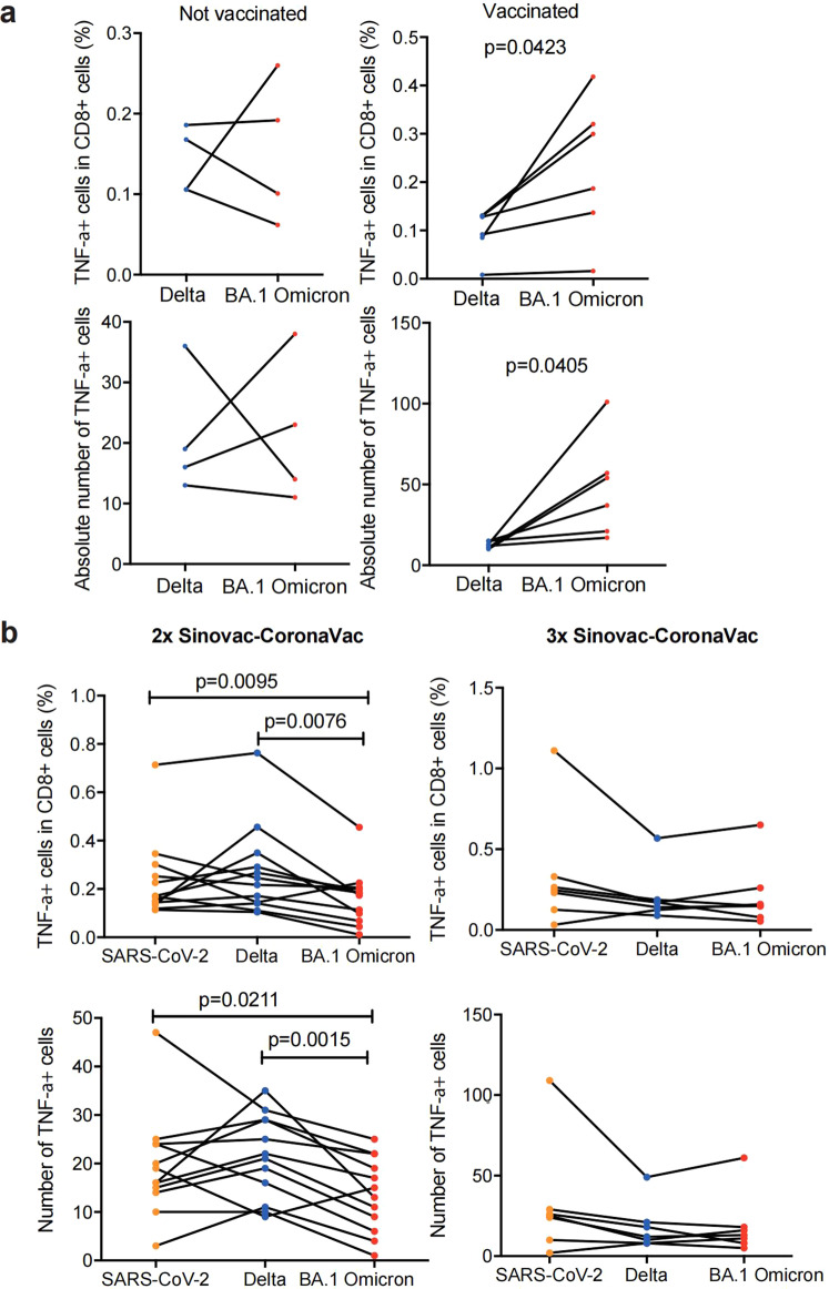
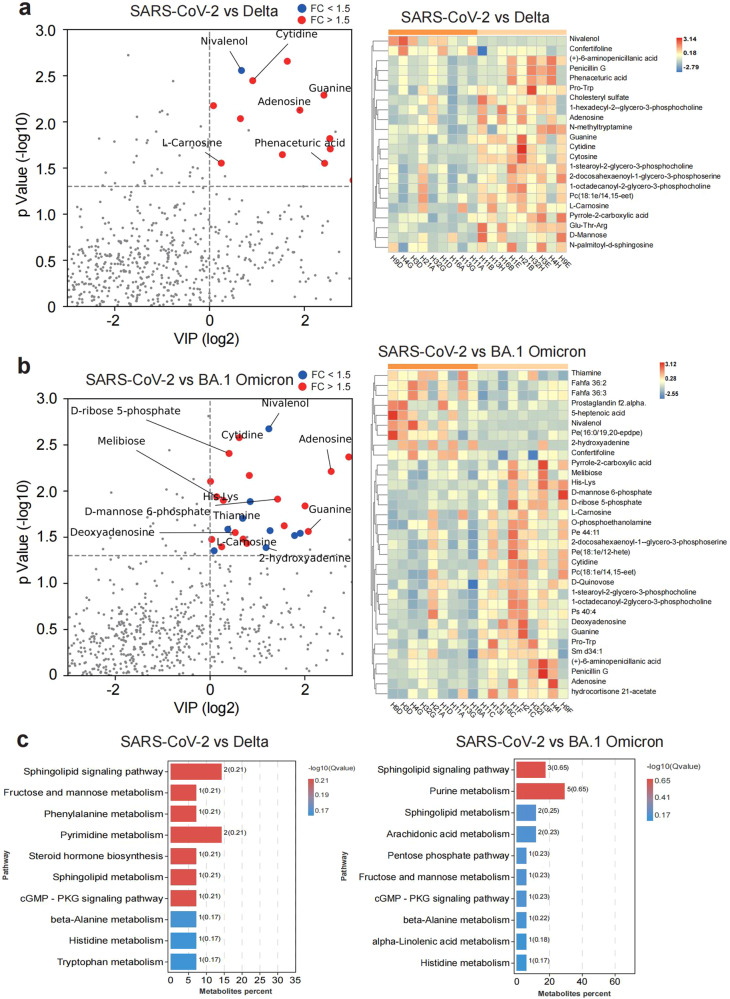
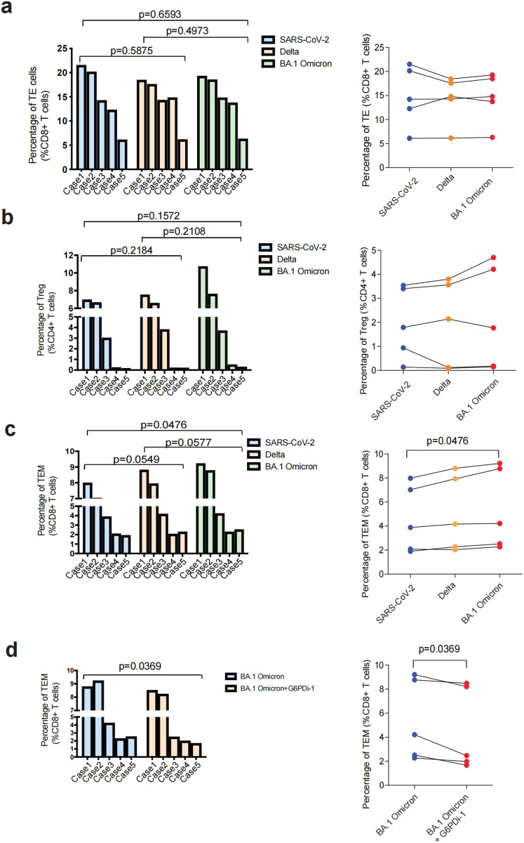
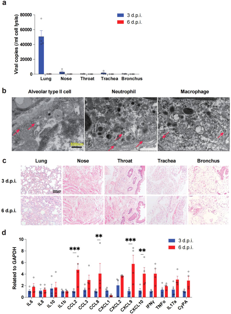
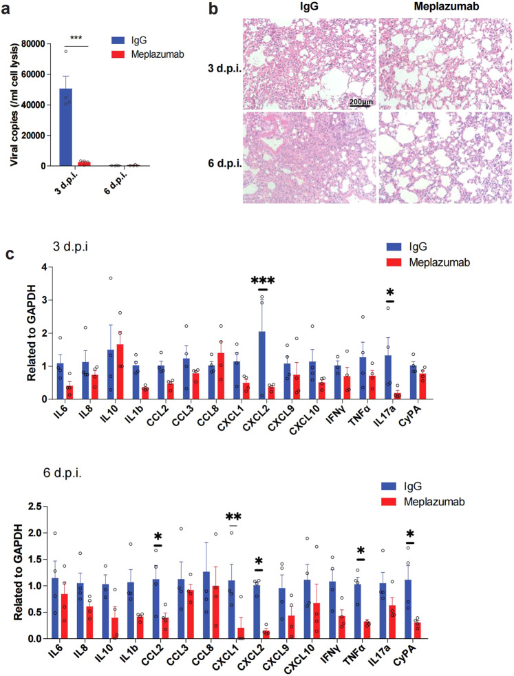
The Omicron variants of SARS-CoV-2, primarily authenticated in November 2021 in South Africa, has initiated the 5th wave of global pandemics. Here, we systemically examined immunological and metabolic characteristics of Omicron variants infection. We found Omicron resisted to neutralizing antibody targeting receptor binding domain (RBD) of wildtype SARS-CoV-2. Omicron could hardly be neutralized by sera of Corona Virus Disease 2019 (COVID-19) convalescents infected with the Delta variant. Through mass spectrometry on MHC-bound peptidomes, we found that the spike protein of the Omicron variants could generate additional CD8 + T cell epitopes, compared with Delta. These epitopes could induce robust CD8 + T cell responses. Moreover, we found booster vaccination increased the cross-memory CD8 + T cell responses against Omicron. Metabolic regulome analysis of Omicron-specific T cell showed a metabolic profile that promoted the response of memory T cells. Consistently, a greater fraction of memory CD8 + T cells existed in Omicron stimulated peripheral blood mononuclear cells (PBMCs). In addition, CD147 was also a receptor for the Omicron variants, and CD147 antibody inhibited infection of Omicron. CD147-mediated Omicron infection in a human CD147 transgenic mouse model induced exudative alveolar pneumonia. Taken together, our data suggested that vaccination booster and receptor blocking antibody are two effective strategies against Omicron.
© 2023. The Author(s).
Conflict of interest statement
The authors declare no competing interests.
Figures
Similar articles
-
Minimal Crossover between Mutations Associated with Omicron Variant of SARS-CoV-2 and CD8+ T-Cell Epitopes Identified in COVID-19 Convalescent Individuals.mBio. 2022 Apr 26;13(2):e0361721. doi: 10.1128/mbio.03617-21. Epub 2022 Mar 1. mBio. 2022. PMID: 35229637 Free PMC article.
-
A Glycosylated RBD Protein Induces Enhanced Neutralizing Antibodies against Omicron and Other Variants with Improved Protection against SARS-CoV-2 Infection.J Virol. 2022 Sep 14;96(17):e0011822. doi: 10.1128/jvi.00118-22. Epub 2022 Aug 16. J Virol. 2022. PMID: 35972290 Free PMC article.
-
The Third dose of CoronVac vaccination induces broad and potent adaptive immune responses that recognize SARS-CoV-2 Delta and Omicron variants.Emerg Microbes Infect. 2022 Dec;11(1):1524-1536. doi: 10.1080/22221751.2022.2081614. Emerg Microbes Infect. 2022. PMID: 35608053 Free PMC article.
-
Pre-Omicron Vaccine Breakthrough Infection Induces Superior Cross-Neutralization against SARS-CoV-2 Omicron BA.1 Compared to Infection Alone.Int J Mol Sci. 2022 Jul 12;23(14):7675. doi: 10.3390/ijms23147675. Int J Mol Sci. 2022. PMID: 35887023 Free PMC article.
-
A mosaic-type trimeric RBD-based COVID-19 vaccine candidate induces potent neutralization against Omicron and other SARS-CoV-2 variants.Elife. 2022 Aug 25;11:e78633. doi: 10.7554/eLife.78633. Elife. 2022. PMID: 36004719 Free PMC article.
Cited by
-
Receptors and Cofactors That Contribute to SARS-CoV-2 Entry: Can Skin Be an Alternative Route of Entry?Int J Mol Sci. 2023 Mar 26;24(7):6253. doi: 10.3390/ijms24076253. Int J Mol Sci. 2023. PMID: 37047226 Free PMC article. Review.
-
Meplazumab, a CD147 antibody, for severe COVID-19: a double-blind, randomized, placebo-controlled, phase 3 clinical trial.Signal Transduct Target Ther. 2025 Apr 14;10(1):119. doi: 10.1038/s41392-025-02208-9. Signal Transduct Target Ther. 2025. PMID: 40222976 Free PMC article. Clinical Trial.
-
SARS-CoV-2-associated lymphopenia: possible mechanisms and the role of CD147.Cell Commun Signal. 2024 Jul 4;22(1):349. doi: 10.1186/s12964-024-01718-3. Cell Commun Signal. 2024. PMID: 38965547 Free PMC article. Review.
-
Prevalence and influencing factors of occupational burnout among healthcare workers in the Chinese mainland during the late 2022 Omicron COVID-19 outbreak: a multicenter cross-sectional study.BMC Public Health. 2025 Jan 15;25(1):171. doi: 10.1186/s12889-024-20930-x. BMC Public Health. 2025. PMID: 39815194 Free PMC article.
-
Timing of general anesthesia for pediatric patients recovering from COVID-19: a prospective cohort study.BMC Anesthesiol. 2024 Jan 2;24(1):11. doi: 10.1186/s12871-023-02390-9. BMC Anesthesiol. 2024. PMID: 38166732 Free PMC article.
References
MeSH terms
Substances
Supplementary concepts
Grants and funding
LinkOut - more resources
Full Text Sources
Medical
Research Materials
Miscellaneous
