. 2023 Jan 21;9(1):16.
doi: 10.1038/s41420-023-01331-2.

Resveratrol protects osteocytes against oxidative stress in ovariectomized rats through AMPK/JNK1-dependent pathway leading to promotion of autophagy and inhibition of apoptosis

Affiliations

Resveratrol protects osteocytes against oxidative stress in ovariectomized rats through AMPK/JNK1-dependent pathway leading to promotion of autophagy and inhibition of apoptosis

Liwei Wei et al. Cell Death Discov. .

Abstract

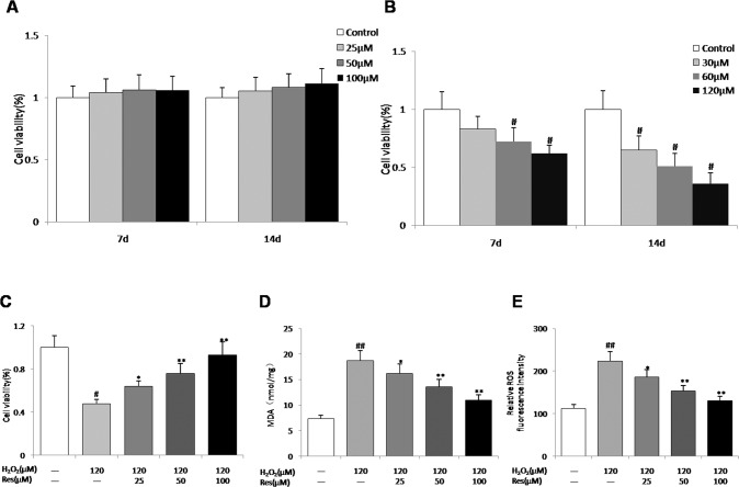
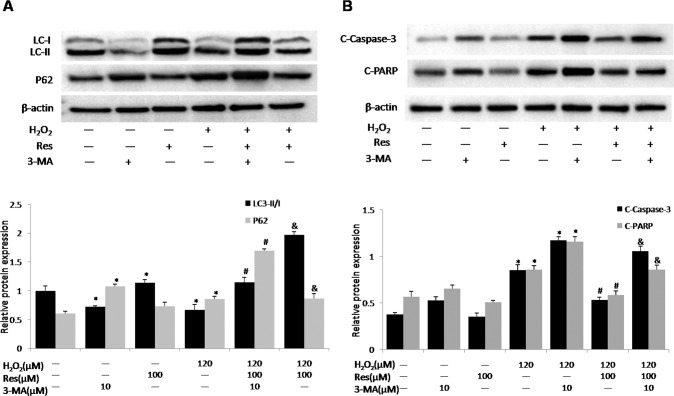
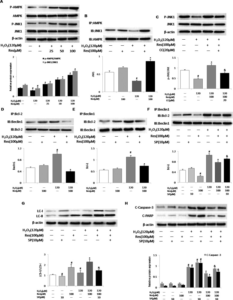
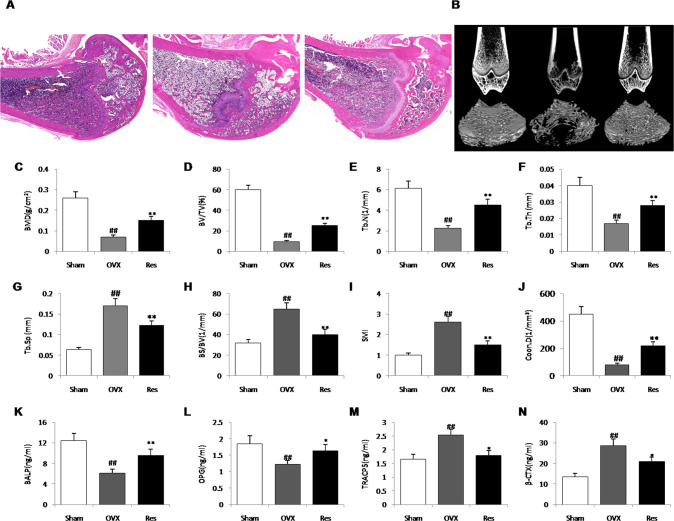
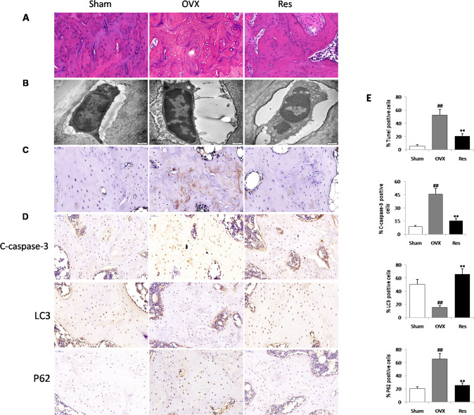
A large number of studies in recent years indicate that osteocytes are the orchestrators of bone remodeling by regulating both osteoblast and osteoclast activities. Oxidative stress-induced osteocyte apoptosis plays critical roles in the pathological processes of postmenopausal osteoporosis. Resveratrol is a natural polyphenolic compound that ameliorates postmenopausal osteoporosis. However, whether resveratrol regulates osteocyte apoptosis via autophagy remains largely unknown. The effects of resveratrol on regulating osteocyte apoptosis and autophagy were analyzed both in vivo and in vitro. In vitro, cultured MLO-Y4 cells were exposed to H2O2 with or without resveratrol. In vivo, an ovariectomy-induced osteoporosis model was constructed in rats with or without daily intraperitoneal injection of 10 mg/kg body weight resveratrol. It was found that resveratrol attenuated H2O2-induced apoptosis through activating autophagy in cultured MLO-Y4 cells, which was mediated by the dissociation of Beclin-1/Bcl-2 complex in AMPK/JNK1-dependent pathway, ultimately regulating osteocytes function. Furthermore, it was shown that resveratrol treatment reduced osteocytes oxidative stress, inhibited osteocytes apoptosis and promoted autophagy in ovariectomized rats. Our study suggests that resveratrol protects against oxidative stress by restoring osteocytes autophagy and alleviating apoptosis via AMPK/JNK1 activation, therefore dissociating Bcl-2 from Beclin-1.


Conflict of interest statement

The authors declare no competing interests.

Figures

Fig. 1
Fig. 1. Efficacy of resveratrol on the viability of MLO-Y4 cells and oxidative stress.
A Cell viability of MLO-Y4 cells following treatment with different concentrations of resveratrol from 7 d to 14 d. B Cell viability of MLO-Y4 cells in different concentrations of H2O2 from 7 d to 14 d. C Cell viability of MLO-Y4 cells administered with resveratrol for 24 h before 120 μM H2O2 administration for 14 d. D, E Effects of different concentrations of resveratrol on MDA and intracellular ROS content in MLO-Y4 cells supernatants after treatment with 120 μM H2O2. n = 6, #P < 0.05 vs control, ##P < 0.01 vs control, *P < 0.05 vs H2O2, **P < 0.01 vs H2O2.
Fig. 2
Fig. 2. Resveratrol inhibits apoptosis and enhances autophagy induced by H2O2 in MLO-Y4 cells.
Cells were exposed to H2O2 (120 μM) and treated with resveratrol (Res, 25, 50, 100 μM) for 14 d. A Representative diagram of annexin V/PI staining. B The apoptotic cells were calculated as the ratio of annexin V + /PI- cells to total cells. C, D The protein expression of Bcl-2, Bax and Caspase-3 in cell lysates were detected by western blotting. E, F MLO-Y4 cells were stained for LC3 (red arrows) and with the nuclear dye DAPI (blue) following the different treatments for 14 d. G, H The protein expression of LC3-II/I and Beclin1 in cell lysates were detected by western blotting. n = 6, #P < 0.05 vs control, ##P < 0.01 vs control, *P < 0.05 vs H2O2, **P < 0.01 vs H2O2.
Fig. 3
Fig. 3. Resveratrol attenuates H2O2-induced apoptosis associated with enhanced autophagy.
MLO-Y4 cells were pretreated with 3-methyladenine (3-MA, 10 μM) for 30 min before exposure to H2O2 (120 μM) and resveratrol (Res, 100 μM) for 14 d. A The protein expression of LC3-II/I and P62 in cell lysates were detected by western blotting. B Apoptosis markers including cleaved caspase-3 (C-caspase-3) and cleaved PARP (C-PARP) in cell lysates were detected by western blotting. n = 3; #P < 0.05 vs. control, *P < 0.05 vs. H2O2, &P < 0.05 vs. H2O2/Res.
Fig. 4
Fig. 4. Resveratrol activates AMPK and JNK1 to disrupt the interactions between Beclin-1 and Bcl-2, thus promoting autophagy and inhibiting apoptosis.
MLO-Y4 cells were pretreated with compound C (CC, 20 μM, AMPK inhibitor), or SP600125 (SP, 10 μM, JNK1 inhibitor) for 30 min before exposure to H2O2 (120 μM). MLO-Y4 Cells were treated with resveratrol for 14 d. A Resveratrol (Res, 25, 50, 100 μM) activated AMPK and JNK1 in MLO-Y4 cells exposed to H2O2. The levels of phosphorylated AMPKɑ (Thr172) and phosphorylation of JNK1 (Thr183/Tyr185) in cells lysates were measured by western blotting. B Resveratrol (Res, 100 μM) enhanced the interaction of AMPKɑ with JNK1 in MLO-Y4 cells exposed to H2O2. The association of AMPKɑ and JNK1 was assessed by immunoprecipitation (IP). C MLO-Y4 cells were pretreated with compound C (CC, 20 μM) for 30 min before exposure to 120 μM H2O2 and treated with resveratrol (Res, 100 μM) for 14 d, JNK1 and phosphorylation of JNK1 (Thr172) protein levels were measured by western blotting. D, E Beclin-1 or Bcl-2 was immunoprecipitated (IP) from cell lysates, and Bcl-2 or Beclin1 in the immunoprecipitate was detected by western blotting. F MLO-Y4 cells were pretreated with SP600125 (SP, 10 μM, JNK1 inhibitor) for 30 min before exposure to H2O2 (120 μM) and treated with resveratrol (Res, 100 μM) for 14 d, The association of Beclin-1 and Bcl-2 was assessed by immunoprecipitation (IP). G, H LC3-II/I protein levels and apoptosis markers (C-Casp3 and C-PARP) in cells lysates were measured by western blotting. n = 3; #P < 0.05 vs. control, *P < 0.05 vs. H2O2, &P < 0.05 vs. H2O2/Res.
Fig. 5
Fig. 5. Protective effect of resveratrol on osteoporosis in ovariectomized rats.
A The hematoxylin-eosin staining in distal femur among groups. B The 2D images of sagittal and transverse section. CJ The trabecula parameters of cancellous bone in the proximal tibial metaphysis static parameters: BMD bone mineral density, BV/TV bone volume/tissue volume, Tb.N trabecular number, Tb.Th trabecular thickness, Tb.Sp trabecular separation, BS/BV Bone surface/bone volume, SMI structure model index, and Conn.D connectivity Density. KN serum bone alkaline phosphatase (BALP), osteoprotegerin (OPG), tartrate resistant acid phosphatase (TRACP-5b), and β-cross-linked c-telopeptide of type I collagen (β-CTX), n = 5, ##P < 0.01 vs. Sham group; *P < 0.05 vs. OVX group and **P < 0.01 vs. OVX group.
Fig. 6
Fig. 6. Effects of resveratrol on oxidative stress status in the ovariectomized model.
AC total antioxidant capacity (T-AOC) (A), catalase (CAT) (B), and superoxide dismutase (SOD) (C) activity in proximal tibias. (D) The protein expression levels of SOD1 and SOD2 were detected by western blot. Data were the means ± SD (n = 6 for each group). ##P < 0.01 vs. Sham group; **P < 0.01 vs. OVX group.
Fig. 7
Fig. 7. Resveratrol administration induced autophagy and inhibited apoptosis of osteocyte in ovariectomized rats.
A Several osteocytes inside the lacunae are observed by hematoxylin-eosin staining. B Ultrastructural architecture of osteocytes in the proximal tibia. C The percentage of apoptotic osteocytes was assessed by Tunel staining in the proximal tibia. D Osteocyte-enriched bone sections were analyzed by immunohistochemistry for Cleaved caspase-3, LC3, P62. E Histomorphometric quantifications of the osteocytes number, ##P < 0.01 vs. Sham group; **P < 0.01 vs. OVX group.
Fig. 8
Fig. 8. Schematic diagram for the molecular regulation of resveratrol against ovariectomized osteoporosis with osteocytes as target.
Resveratrol protects osteocytes by regulating the switch between autophagy and apoptotic machinery under estrogen deficiency conditions, which is attributed by AMPK/JNK-mediated dissociation of Beclin-1-Bcl-2. ROS reactive oxygen species, AMPK Adenosine 5‘-monophosphate (AMP)-activated protein kinase, JNK1 c-Jun N-terminal kinase1.

Similar articles

Cited by

References

    1. Farr JN, Rowsey JL, Eckhardt BA, Thicke BS, Fraser DG, Tchkonia T, et al. Independent roles of estrogen deficiency and cellular senescence in the pathogenesis of osteoporosis: evidence in young adult mice and older humans. J Bone Miner Res. 2019;34:1407–18. doi: 10.1002/jbmr.3729. - DOI - PMC - PubMed
    1. Watson SL, Weeks BK, Weis LJ, Harding AT, Horan SA, Beck BR. High-intensity resistance and impact training improves bone mineral density and physical function in postmenopausal women with osteopenia and osteoporosis: The LIFTMOR randomized controlled trial. J Bone Miner Res. 2018;33:211–20. doi: 10.1002/jbmr.3284. - DOI - PubMed
    1. Raggatt LJ, Partridge NC. Cellular and molecular mechanisms of bone remodeling. J Biol Chem. 2010;285:25103–8. doi: 10.1074/jbc.R109.041087. - DOI - PMC - PubMed
    1. Plotkin LI, Bellido T. Osteocytic signalling pathways as therapeutic targets for bone fragility. Nat Rev Endocrinol. 2016;12:593–605. doi: 10.1038/nrendo.2016.71. - DOI - PMC - PubMed
    1. Qin L, Liu W, Cao H, Xiao G. Molecular mechanosensors in osteocytes. Bone Res. 2020;8:23. doi: 10.1038/s41413-020-0099-y. - DOI - PMC - PubMed