. 2023 Jan 21;13(1):1221.
doi: 10.1038/s41598-023-28352-2.

Identification of platelet subpopulations in cryopreserved platelet components using multi-colour imaging flow cytometry

Affiliations

Identification of platelet subpopulations in cryopreserved platelet components using multi-colour imaging flow cytometry

Lacey Johnson et al. Sci Rep. .

Abstract

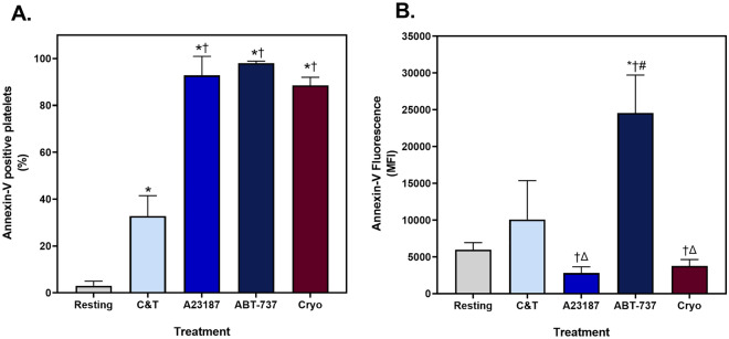
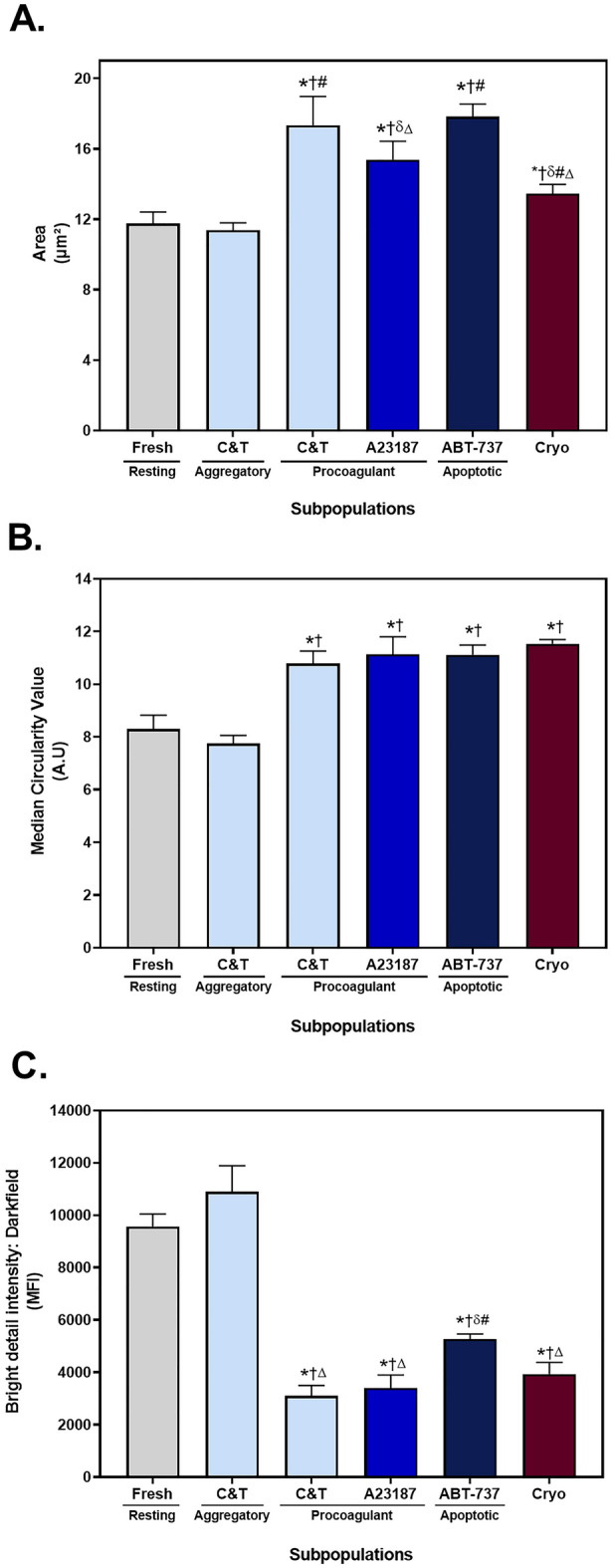
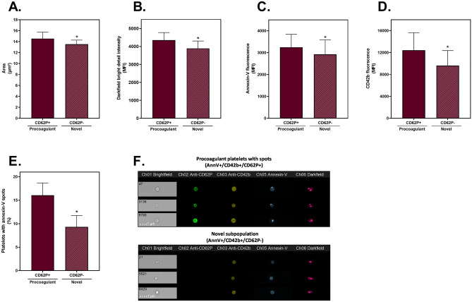
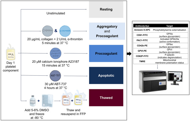
Cryopreservation of platelets, at - 80 °C with 5-6% DMSO, results in externalisation of phosphatidylserine and the formation of extracellular vesicles (EVs), which may mediate their procoagulant function. The phenotypic features of procoagulant platelets overlap with other platelet subpopulations. The aim of this study was to define the phenotype of in vitro generated platelet subpopulations, and subsequently identify the subpopulations present in cryopreserved components. Fresh platelet components (n = 6 in each group) were either unstimulated as a source of resting platelets; or stimulated with thrombin and collagen to generate a mixture of aggregatory and procoagulant platelets; calcium ionophore (A23187) to generate procoagulant platelets; or ABT-737 to generate apoptotic platelets. Platelet components (n = 6) were cryopreserved with DMSO, thawed and resuspended in a unit of thawed plasma. Multi-colour panels of fluorescent antibodies and dyes were used to identify the features of subpopulations by imaging flow cytometry. A combination of annexin-V (AnnV), CD42b, and either PAC1 or CD62P was able to distinguish the four subpopulations. Cryopreserved platelets contained procoagulant platelets (AnnV+/PAC1-/CD42b+/CD62P+) and a novel population (AnnV+/PAC1-/CD42b+/CD62P-) that did not align with the phenotype of aggregatory (AnnV-/PAC1+/CD42b+/CD62P+) or apoptotic (AnnV+/PAC1-/CD42b-/CD62P-) subpopulations. These data suggests that the enhanced haemostatic potential of cryopreserved platelets may be due to the cryo-induced development of procoagulant platelets, and that additional subpopulations may exist.


Conflict of interest statement

The authors declare no competing interests.

Figures

Figure 1
Figure 1
Representative scatterplots and images of platelets and extracellular vesicles. Fresh components were either unstimulated (resting), or stimulated with collagen and thrombin (C&T), A23187 or ABT-737 (n = 6 for each treatment). Cryopreserved (cryo) platelet components were thawed and reconstituted in plasma (n = 6). The populations of platelets (PLTs) and extracellular vesicles (EVs) were distinguished based on brightfield area and aspect ratio intensity. (A) A representative density scatterplot of each group. Representative brightfield images of (B) platelets, (C) extracellular vesicles and (D) ungated events. (E) The mean + SD (error bars) CD61+ EVs (% of sample) in each group. * Indicates p < 0.05 compared to resting; † Indicates p < 0.05 compared to C&T; # Indicates p < 0.05 compared to A23187; Δ Indicates p < 0.05 compared to ABT-737.
Figure 2
Figure 2
The proportion of platelets with externalised phosphatidylserine. Fresh components were either unstimulated (resting), or stimulated with collagen and thrombin (C&T), A23187 or ABT-737 (n = 6 for each treatment). Cryopreserved (cryo) platelet components were thawed and reconstituted in plasma (n = 6). Samples were analysed by imaging flow cytometry, with 7500 platelet events recorded. (A) The proportion of annexin-V positive (AnnV+) platelets and (B) the median fluorescence intensity (MFI) of AnnV+ platelets was determined from the APC fluorescence channel (Ch 05). The data represent the mean + SD (error bars). * Indicates p < 0.05 compared to resting; † Indicates p < 0.05 compared to the C&T; # Indicates p < 0.05 compared to A23187; Δ indicates p < 0.05 compared to ABT-737.
Figure 3
Figure 3
Representative images of the phenotype of platelet subpopulations within each treatment group. Fresh components were either unstimulated (resting), or stimulated with collagen and thrombin (C&T), A23187 or ABT-737 (n = 6 for each treatment). Cryopreserved platelet components were thawed and reconstituted in plasma (n = 6). The platelets were stained with four panels containing three different fluorescent antibodies/dyes and analysed by imaging flow cytometry, with 7500 platelet events recorded. Representative images of the phenotype of the most prominent platelet subpopulation(s) in the treatment group are presented. The pattern of staining observed in (A) resting platelets was annexin-V (AnnV)/CD61+/TMRE+/PAC1/GPVI+/CD62P/CD42b+, (B) aggregatory platelets (from C&T stimulation) were AnnV/CD61+/TMRE+/PAC1+/GPVI+/CD62P+/CD42b+, (C) procoagulant platelets (from C&T stimulation) were AnnV+/CD61+/TMRE/PAC1/GPVI/CD62P+/CD42b+, (D) procoagulant platelets (from A23187 stimulation) were AnnV+/CD61+/TMRE/PAC1/GPVI/CD62P+/CD42b+, (E) apoptotic platelets were AnnV+/CD61+/TMRE/PAC1/GPVI/CD62P/CD42b, and (F) two populations of cryopreserved platelets were observed: AnnV+/CD61+/TMRE/PAC1/GPVI/CD62P+/CD42b+ and AnnV+/CD61+/TMRE/PAC1/GPVI/CD62P/CD42b+.
Figure 4
Figure 4
Morphological parameters of platelet subpopulations. Fresh components were either unstimulated (resting), or stimulated with collagen and thrombin (C&T), A23187 or ABT-737 (n = 6 for each treatment). Cryopreserved (cryo) platelet components were thawed and reconstituted in plasma (n = 6). Samples were analysed by imaging flow cytometry, with 7500 platelet events recorded. (A) The median area was calculated using a custom mask (combining the brightfield and fluorescent marker channels). (B) The circularity of platelets was determined using a custom mask (combining the brightfield and fluorescent marker channels). (C) The internal complexity of the platelets, as indicated by the median intensity of darkfield bright detail was determined using a custom mask (combining brightfield, fluorescent and darkfield channels). The data represent the mean + SD (error bar). * Indicates p < 0.05 compared to resting; † Indicates p < 0.05 compared to the aggregatory phenotype derived from C&T stimulation; δ Indicates p < 0.05 compared to the procoagulant phenotype derived from C&T stimulation; # Indicates p < 0.05 compared to A23187; Δ Indicates p < 0.05 compared to ABT-737.
Figure 5
Figure 5
Interrogation of the CD62P+ and CD62P subpopulations identified in cryopreserved platelets. Cryopreserved platelet components were thawed and reconstituted in plasma (n = 6). Samples were analysed by imaging flow cytometry, with 7500 platelet events recorded. The annexin-V positive (AnnV+)/CD42b+ subpopulations which were either CD62P+ (procoagulant) or CD62P (novel) were compared. (A) The median area was calculated using a custom mask (combining the brightfield and fluorescent marker channels). (B) The median intensity of darkfield bright detail was determined using a custom mask (combining brightfield, fluorescent and darkfield channels). The median fluorescence intensity (MFI) of (C) AnnV and (D) CD42b of the positive platelets. (E) The proportion of each subpopulation containing AnnV+ spots was calculated using the spot count feature, with (F) representative images of CD62P+ and CD62P cryopreserved platelets displaying the presence and absence of AnnV+ spots. The data represent the mean + SD (error bar). * Indicates p < 0.05 compared to CD62P+ cryopreserved platelets.
Figure 6
Figure 6
The experimental design for generation of platelet subpopulations. Fresh platelet components were either unstimulated (resting) or stimulated with collagen and α-thrombin (C&T), calcium ionophore A23187 or ABT-737 (n = 6 for each treatment). Stimulation with these agents is known to induce aggregatory and procoagulant (C&T), procoagulant (A23187) or apoptotic (ABT-737) platelets, respectively. Fresh platelet components (n = 6) were frozen at  − 80 °C with 5–6% DMSO. Prior to testing, cryopreserved platelet components were thawed at 37 °C and reconstituted in fresh frozen plasma (FFP). Samples from each group were stained with multiple fluorescent markers, as indicated, and data were acquired using an imaging flow cytometer (ImageStreamX Mark II) and then analysed using IDEAS software.

Similar articles

Cited by

References

    1. Kaufman RM, et al. Platelet transfusion: A clinical practice guideline from the AABB. Ann. Intern. Med. 2015;162:205–213. doi: 10.7326/m14-1589. - DOI - PubMed
    1. Lelkens CC, Koning JG, de Kort B, Floot IB, Noorman F. Experiences with frozen blood products in the Netherlands military. Transfus. Apher. Sci. 2006;34:289–298. doi: 10.1016/j.transci.2005.11.008. - DOI - PubMed
    1. Valeri CR, Ragno G, Khuri S. Freezing human platelets with 6 percent dimethyl sulfoxide with removal of the supernatant solution before freezing and storage at − 80 degrees C without postthaw processing. Transfusion. 2005;45:1890–1898. doi: 10.1111/j.1537-2995.2005.00647.x. - DOI - PubMed
    1. Johnson LN, Winter KM, Reid S, Hartkopf-Theis T, Marks DC. Cryopreservation of buffy-coat-derived platelet concentrates in dimethyl sulfoxide and platelet additive solution. Cryobiology. 2011;62:100–106. doi: 10.1016/j.cryobiol.2011.01.003. - DOI - PubMed
    1. Johnson L, Reade MC, Hyland RA, Tan S, Marks DC. In vitro comparison of cryopreserved and liquid platelets: Potential clinical implications. Transfusion. 2015;55:838–847. doi: 10.1111/trf.12915. - DOI - PubMed

Publication types

Substances