. 2023 Jan 21;6(1):79.
doi: 10.1038/s42003-022-04402-9.

Single-cell transcriptomics reveal extracellular vesicles secretion with a cardiomyocyte proteostasis signature during pathological remodeling

Affiliations

Single-cell transcriptomics reveal extracellular vesicles secretion with a cardiomyocyte proteostasis signature during pathological remodeling

Eric Schoger et al. Commun Biol. .

Erratum in

Abstract

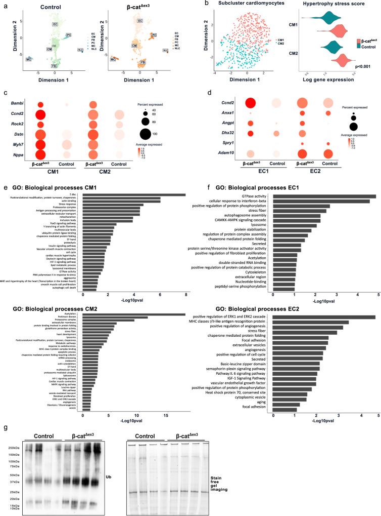
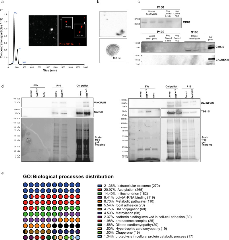
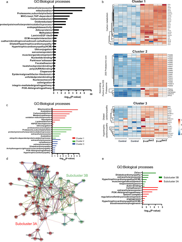
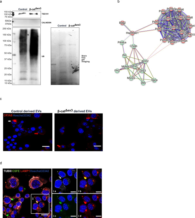
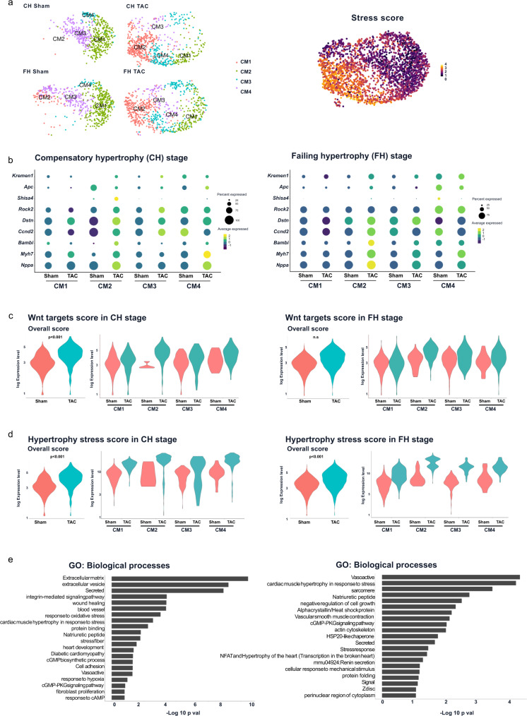
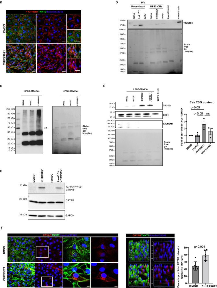
Aberrant Wnt activation has been reported in failing cardiomyocytes. Here we present single cell transcriptome profiling of hearts with inducible cardiomyocyte-specific Wnt activation (β-catΔex3) as well as with compensatory and failing hypertrophic remodeling. We show that functional enrichment analysis points to an involvement of extracellular vesicles (EVs) related processes in hearts of β-catΔex3 mice. A proteomic analysis of in vivo cardiac derived EVs from β-catΔex3 hearts has identified differentially enriched proteins involving 20 S proteasome constitutes, protein quality control (PQC), chaperones and associated cardiac proteins including α-Crystallin B (CRYAB) and sarcomeric components. The hypertrophic model confirms that cardiomyocytes reacted with an acute early transcriptional upregulation of exosome biogenesis processes and chaperones transcripts including CRYAB, which is ameliorated in advanced remodeling. Finally, human induced pluripotent stem cells (iPSC)-derived cardiomyocytes subjected to pharmacological Wnt activation recapitulated the increased expression of exosomal markers, CRYAB accumulation and increased PQC signaling. These findings reveal that secretion of EVs with a proteostasis signature contributes to early patho-physiological adaptation of cardiomyocytes, which may serve as a read-out of disease progression and can be used for monitoring cellular remodeling in vivo with a possible diagnostic and prognostic role in the future.


Conflict of interest statement

The authors declare no competing interests.

Figures

Fig. 1
Fig. 1. Dysregulated genes and processes in cardiomyocytes and endothelial cells from β-catΔex3 hearts.
a Representative uniform manifold approximation and projection (UMAP) plot after scRNA-seq and data integration of cardiac cells isolated from control (673 cells) or β-catΔex3 (601 cells) hearts. Six cell clusters were identified and exclusively classified into cardiomyocytes (CM), endothelial cells (EC), fibroblast (FB), pericytes (PC), macrophage (MC) and neural-like cells (NLC) in both conditions. b UMAP plot of cardiomyocyte subclusters CM1, CM2) with respective hypertrophic stress scores as defined by the expression of Acta1, Nppa, Nppb and Ankrd1. c Dot plot depicting expression of selected cardiac stress and Wnt signaling target transcripts in cardiomyocytes from β-catΔex3 and control hearts as indicated. d Selected top categories from GO biological processes enrichment of upregulated DEGs in CM1 and CM2 of β-catΔex3 representing transcriptional responses. e Dot plot depicting expression of selected developmental and angiogenesis transcripts in endothelial cells (EC1 and EC2) from β-catΔex3 and control hearts as indicated. f Selected top categories from GO biological processes enrichment of upregulated DEGs in EC1 and EC2 of β-catΔex3 representing transcriptional responses in endothelial clusters. GO enrichment represents –log10 p-value (p-value <0.05) and term fusion was applied. g Increased Ub-protein abundance in β-catΔex3 hearts versus control. Staining-free gel is provided as loading control.
Fig. 2
Fig. 2. Increased cardiac EV cargo secretion in β-catΔex3 hearts.
a Nanoparticle tracking analyses (NTA) confirmed isolation of extracellular vesicles with a mean size of 160.0 ± 69 nm. Visualization of the purified vesicles by hydrophilic fluorescence analog of cholesterol (Chol-PEG-KK114) staining. b Electron microscopy confirmed the isolation of round and cup-shaped particles, typical for exosomal vesicles less than 200 nm in size. c Western blot showing expression of the exosome marker CD81 in the higher speed-centrifugation fraction (P100) from mouse hearts and the absence of Golgi-marker GM130 and endoplasmic reticulum marker Calnexin, which were present in the cell pellets and lower-speed centrifugation (S100). d Western blot showing increased exosomal marker TSG101 in EV isolated from β-catΔex3 hearts versus control, as well as showing absence of Calnexin, GAPDH and Vinculin expression in both conditions upon equally amount of loaded proteins as visualized by the stain free gel image. e GO biological processes distribution of enrichment analysis of total proteomic data (573 proteins) obtained from isolated vesicles of β-catΔex3 hearts and control confirming an association of vesicle contained proteins with “extracellular exosomes” (n = 2 (each a pool of 2-3 hearts) biological replicates and technical triplicates for NTA and MS analysis, n = 4, biological replicates (for Western blots). GO enrichment represents –log10 p-value (p-value <0.05) and term fusion was applied.
Fig. 3
Fig. 3. Stress-related proteome signature in cardiac exosomes from β-catΔex3 hearts.
a GO biological processes distribution of enrichment analysis of significantly enriched proteins identified in isolated vesicles from β-catΔex3 hearts versus control. b Heatmap of cluster analysis using STRING functional annotation protein interaction database on the predicted enriched loading proteins in exosomes from β-catΔex3 tissue using the K-means algorithm. c GO biological processes analysis of the three different clusters identified: Cluster 1 (metabolism); Cluster 2 (proteasome associated proteins) and Cluster 3 (cardiomyopathy associated proteins). c, d Further subclassification of the Cluster 3 in sub-clusters 3A and 3B and (e) associated GO biological processes. GO enrichment represents –log10 p-value (p-value <0.05) and term fusion was applied.
Fig. 4
Fig. 4. Ub-proteasomal-related proteome signature in exosomes from β-catΔex3 cardiomyocytes.
a Increased Ub-protein abundance in β-catΔex3 hearts versus control. Stain-free gel is provided as loading control. b Protein-protein interaction analysis using STRING functional annotation database on the predicted enriched proteasome, chaperones and cardiac proteins identified in β-catΔex3 exosomes including CRYAB. c Representative confocal images of immunofluorescence stainings of Neuro2a cells that were exposed to β-catΔex3 and control derived-cardiac exosomes and stained for CRYAB. Perinuclear staining (red arrows) and increased protein accumulation (white arrows) was observed in recipient cells (n = 3, technical replicates). d Representative confocal images of immunofluorescence staining of Neuro2a cells that were exposed to carboxyfluorescein succinimidyl ester (CSFE)-ex vivo labeled exosomes from β-catΔex3 hearts, stained with Tubulin III (TUBIII) and LAMP1 (n = 3, technical replicates). The white box (i) indicates the region that is depicted at the right side showing the different stainings (ia-id). Hoechst33342 was used for nucleus visualization. Scale bar = 20 µm.
Fig. 5
Fig. 5. Differential EV-signaling in cardiomyocyte populations upon hypertrophic remodeling.
a UMAP plot of unbiased reclustering of the cardiomyocyte cluster identified after scRNA-seq and data integration. Cells were isolated from control (sham) and hypertrophic (transaortic constriction (TAC)) hearts at an early compensatory (CH) and late failing (FH) stages (average of 650 cells per condition and stage). Four sub-clusters were identified (CM1-CM4). Corresponding hypertrophic stress scores as defined by the expression of Acta1, Nppa, Nppb and Ankrd1 are depicted for all cells. b Representative dot plots showing expression levels for the stress genes Myh7, Nppa, cardiac Wnt-target genes Dstn, Rock2, Bambi, Ccnd2 and Wnt inhibitors Kremmen1, Shisa4, Apc in clusters CM1-CM4 from sham and TAC hearts of CH (left) and FH (right) stages. Violin plots showing Wnt signaling target and stress scores in the different CM clusters for CH (c) and FH (d) stages. e Selected top categories from GO biological processes enrichment of overlapping DEGs obtained from sham vs TAC comparison in CH (left) and FH (right). GO enrichment represents –log10 p-value (p-value <0.05) and term fusion was applied.
Fig. 6
Fig. 6. Increased exosomal and hypoxic scores in cardiomyocyte populations upon hypertrophic remodeling.
Representative violin plots showing expression levels for the cardiac chaperone Cryab and exosomal makers Cd9, Cd81 and Cd63 in clusters CM1-CM4 from sham (pink) and TAC (orange) hearts of CH (a) and FH (b). Hypoxia (c) and exosomal scores (d) in sham and TAC cardiomyocytes in CH and FH.
Fig. 7
Fig. 7. Increased exosomal cargo release in human iPSC-derived cardiomyocytes upon Wnt activation.
a Confocal immunofluorescence images showing nuclear translocation and accumulation of the transcriptionally active P-Ser675-CTNNB1 in TNNT2-positive iPSC-derived cardiomyocytes upon CHIR99021 (CHIR) treatment validating WNT/β-catenin signaling induction versus DMSO control-treated cells. Western blot showing increased (b) TSG101 levels and (c) Ubiquitinated protein abundance in exosomal preparations from CHIR-treated iPSC-derived cardiomyocytes versus control (DMSO), (TGFβ1 was included as additional stress control in b) upon equal amounts of loaded proteins as visualized by the stain free gel image (biological replicates n = 3 (b) and n = 2 (c) technical replicates). In b exosome preparation from β-catΔex3 and control hearts were included for direct comparison. d Western blot and corresponding quantification showing increased TSG101 levels in exosomal preparations from CHIR-treated iPSC-derived cardiomyocytes versus control (DMSO) and a partial rescue upon concomitant treatment with Iso-Quercetin (Iso QC), blocking Wnt transcriptional activity. e Western blot showing transcriptionally active CTNNB1 and CRYAB expression in cell lysates from CHIR, Iso QC and CHIR/Iso QC treated iPSC-derived cardiomyocytes. f Confocal immunofluorescence images showing cytosolic accumulation of CRYAB in CHIR-treated iPSC-derived cardiomyocytes. Hoechst33342 was used for nucleus visualization and corresponding semiquantification is depicted, which is showing the intensity of CRYAB staining normalized to the total TNNT2 intensity (n = 6, technical replicates, biological duplicates). Scale bar = 20 µm. Data are shown as mean ± SEM; unpaired Student’s t test.
Fig. 8
Fig. 8. Scheme summarizing the finding in this study.
Our study revealed an increased EV-cargo secretion including Z-disk proteins prone to misfolding (DES, TNN), proteasome components and chaperone associated proteins by cardiomyocytes upon Wnt activation and pressure overload induced stress. This was accompanied by a concomitant activation of hypoxia response, which is known to activate EV-mediated processes. This response was more accentuated in early compensatory hypertrophic remodeling, suggesting their contribution to hypertrophic disease adaptation, and may overlap with the endosomal autophagic pathway at the formation of amphisomes, also activated upon stress. It is tempting to speculate that increased cellular stress induces protein misfolding, which triggers an excess of ubiquitination and CRYAB-mediated processes activation and the amphisomes are redirected to the exosomal release. Created with BioRender.com.

References

    1. Kumar S, et al. Hypoxia-induced mitogenic factor promotes cardiac hypertrophy via calcium-dependent and hypoxia-inducible factor-1alpha mechanisms. Hypertension. 2018;72:331–342. doi: 10.1161/HYPERTENSIONAHA.118.10845. - DOI - PubMed
    1. Vujic A, Natarajan N, Lee RT. Molecular mechanisms of heart regeneration. Semin. Cell Developmental Biol. 2020;100:20–28. doi: 10.1016/j.semcdb.2019.09.003. - DOI - PMC - PubMed
    1. Iyer LM, et al. A context-specific cardiac beta-catenin and GATA4 interaction influences TCF7L2 occupancy and remodels chromatin driving disease progression in the adult heart. Nucleic Acids Res. 2018;46:2850–2867. doi: 10.1093/nar/gky049. - DOI - PMC - PubMed
    1. Hou, N. et al. Transcription factor 7-like 2 mediates canonical Wnt/beta-catenin signaling and c-Myc upregulation in heart failure. Circ. Heart Fail.9, 10.1161/CIRCHEARTFAILURE.116.003010 (2016). - PMC - PubMed
    1. Clevers H, Loh KM, Nusse R. An integral program for tissue renewal and regeneration: Wnt signaling and stem cell control. Science. 2014;346:1248012. doi: 10.1126/science.1248012. - DOI - PubMed

Publication types