. 2023 Jan 22;11(1):18.
doi: 10.1186/s40478-023-01513-0.

NAD salvage pathway machinery expression in normal and glaucomatous retina and optic nerve

Affiliations

NAD salvage pathway machinery expression in normal and glaucomatous retina and optic nerve

James R Tribble et al. Acta Neuropathol Commun. .

Abstract

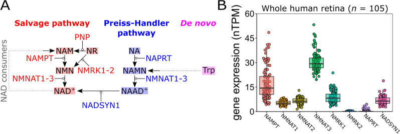
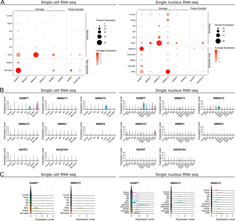
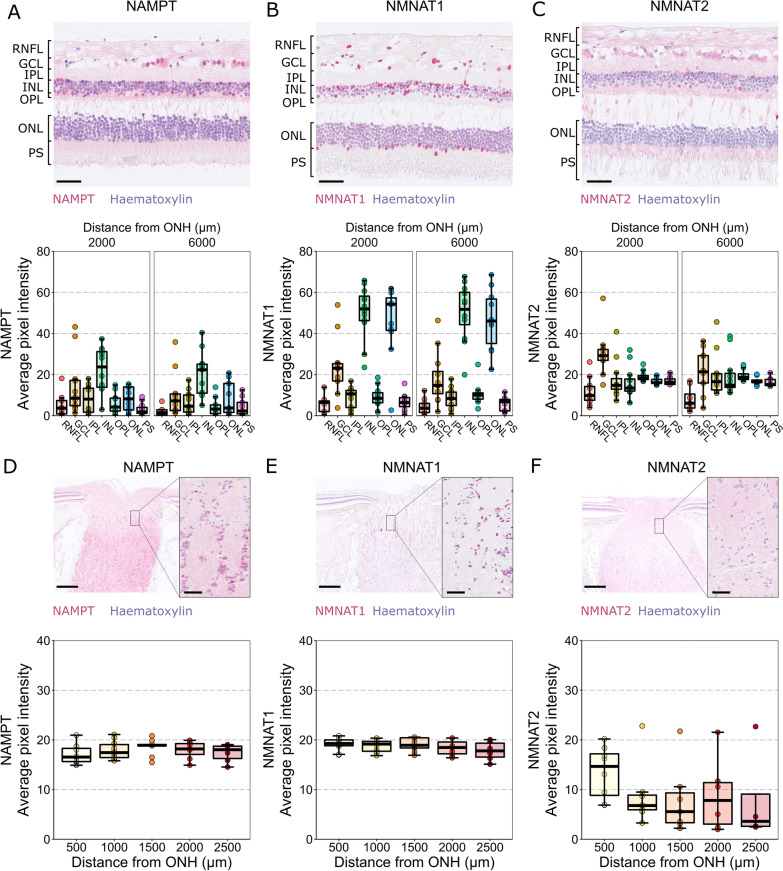
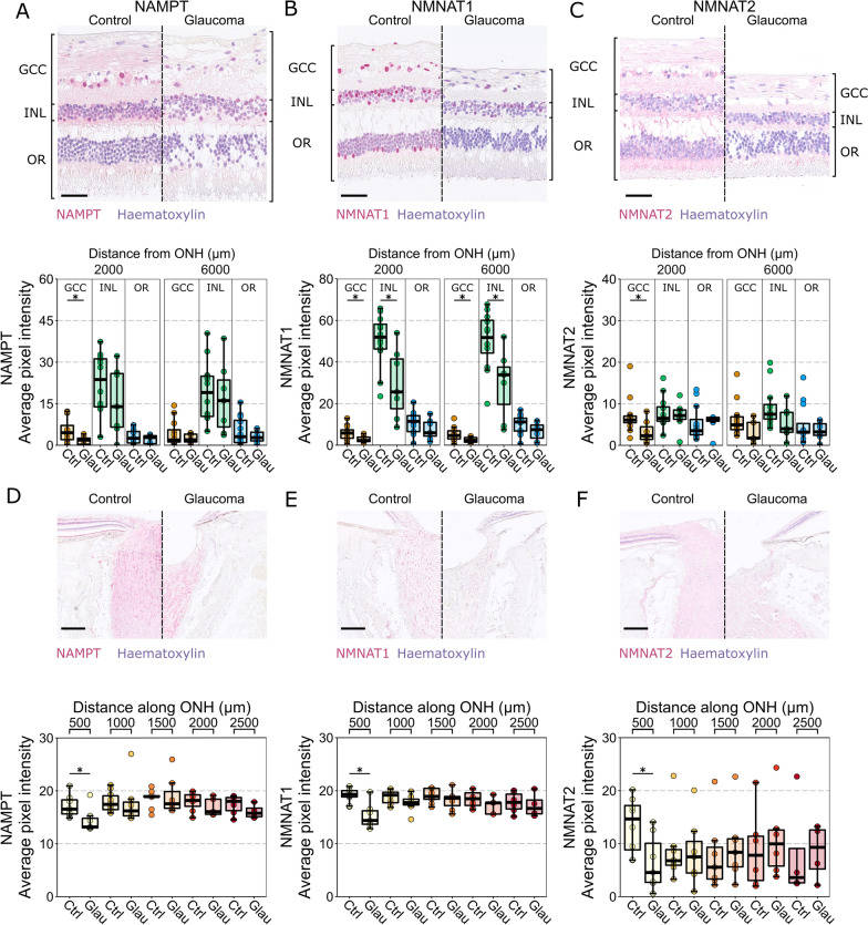
Glaucoma is the leading cause of irreversible blindness and is a major health and economic burden. Current treatments do not address the neurodegenerative component of glaucoma. In animal models of glaucoma, the capacity to maintain retinal nicotinamide adenine dinucleotide (NAD) pools declines early during disease pathogenesis. Treatment with nicotinamide, an NAD precursor through the NAD salvage pathway, robustly protects against neurodegeneration in a number of glaucoma models and improves vision in existing glaucoma patients. However, it remains unknown in humans what retinal cell types are able to process nicotinamide to NAD and how these are affected in glaucoma. To address this, we utilized publicly available RNA-sequencing data (bulk, single cell, and single nucleus) and antibody labelling in highly preserved enucleated human eyes to identify expression of NAD synthesizing enzyme machinery. This identifies that the neural retina favors expression of the NAD salvage pathway, and that retinal ganglion cells are particularly enriched for these enzymes. NMNAT2, a key terminal enzyme in the salvage pathway, is predominantly expressed in retinal ganglion cell relevant layers of the retina and declines in glaucoma. These findings suggest that human retinal ganglion cells can directly utilize nicotinamide and could maintain a capacity to do so in glaucoma, showing promise for ongoing clinical trials.

Keywords: Axon degeneration; Glaucoma; Metabolism; NAD; Neurodegeneration; Nicotinamide; Optic nerve; Retinal ganglion cell.


Conflict of interest statement

PAW is an inventor on an awarded US patent held by The Jackson Laboratory for nicotinamide treatment in glaucoma (“Treatment and prevention of ocular neurodegenerative disorder”, US11389439B2). All other authors declare that they have no competing interests.

Figures

Fig. 1
Fig. 1
NAD synthesis pathways are represented in the retina. A NAD is synthesized through 3 pathways: the NAD salvage pathway, the Preiss-Handler pathway, and de novo from tryptophan. The salvage pathway (red) converts nicotinamide (NAM, a main dietary from of vitamin B3; also known as niacinamide) to NAD+ in a two-step reaction. NAM is converted to nicotinamide mononucleotide (NMN) by the enzyme NAMPT, which is then converted to NAD+ by NNMNATs (isoforms 1–3 are expressed in different cells and localize to different cellular compartments). Alternatively, nicotinamide riboside (NR) can be converted to either NAM (by the enzyme PNP) or NMN (by the enzyme NMRK, isoforms 1–2). NAD+ is recycled to NAM by NAD consumers, allowing cells to replenish NAD without constant influx of dietary precursors. The Preiss-Handler pathway (blue) converts nicotinic acid (NA; also known as niacin) to NAD+ in a three-step reaction. NA is converted to nicotinic acid mononucleotide (NAMN) by the enzyme NAPRT, which is converted to nicotinic acid adenine dinucleotide (NAAD+) by NNMNATs, and finally to NAD+ by the enzyme NADSYN1. Tryptophan can be converted to NAD+ through de novo synthesis (magenta) involving the kinurenine pathway (a six-step reaction which generates NAMN). Enzymes are shown as gene names (HGNC). B Gene expression of NAD-synthesizing enzymes was examined in publicly available bulk sequenced mRNA in 105 whole human retina. NAD-salvage pathway transcripts are well expressed (expect for the NMRK2 isoform). NAPRT in the Preiss-Handler pathway is lowly expressed, whereas NADSYN1 is well expressed suggesting that the retina may favor the NAD-salvage pathway
Fig. 2
Fig. 2
Retinal neurons predominantly express NAD-salvage pathway transcripts. A We examined independent datasets from single cell RNA-sequencing of normal human retina (left) and single nucleus RNA-sequencing of normal human retina (right). Expression of transcripts encoding NAD synthesizing enzyme machinery was compared across cell types of the retina. Retinal neurons demonstrate greater expression levels in a higher proportion of cells for the NAD-salvage pathway (NAMPT, NMNAT1-3) than the Preiss-Handler Pathway (NAPRT, NADSYN1). NAMPT expression is greater than NMRK1-2, suggesting a favoring of nicotinamide as an NAD-salvage pathway substrate over the alternative nicotinamide riboside. Glia and other non-neuronal support cells of the retina also favor the NAD-salvage pathway but have a greater relative expression of Preiss-Handler Pathway and NMRK1 than neurons. B NAMPT, NMNAT1, and NMNAT2 were expressed to a greater extent in RGCs over other retinal neurons. Of the non-neuronal cell types, only microglia/myeloid cells and RPE cells demonstrated strong expression of NAD synthesizing enzyme machinery. C For most cell types, when considering genes with high expression, the distribution of expression within cell types appeared normal, as demonstrated by ridge plots. In the single cell RNA-sequencing (left), distribution of NAMPT and NMNAT2 suggest the possibility of distinct populations, perhaps reflecting different RGC subtypes. This was not observed in the single nucleus sequencing, where distribution of expression appeared more normal for all cell types, with the exception of NMNAT2 in RGCs which had a greater variance, and expression of NAMPT and NMNAT3 in from RPE cells. Retinal neurons: cones, rods, horizontal cells (HCs), bipolar cells (BPs), amacrine cells (ACs), and retinal ganglion cells (RGCs). Non-neuronal retinal cells: myeloid/microglia, Müller glia, astrocytes, vascular cells, and retinal pigment epithelial cells (RPE cells)
Fig. 3
Fig. 3
Labelling of NAD salvage pathway machinery in human retina. A NAMPT labelling is detected across all retinal layers, localizing to both nuclear (haematoxylin + ve) and cytoplasmic (haematoxylin −ve) cellular compartments, with the most intense labeling in the INL (n = 11 eyes). B NMNAT1 labelling is strongest in nuclear layers (GCL, INL, ONL) localizing to nuclei (n = 11 eyes). C NMNAT2 labelling localized to cytoplasmic compartments and was detected across all retinal layers, with the greatest intensity in the GCL, reflecting high expression in RGCs (n = 11 eyes). D In the ONH, NAMPT followed the same trend as in the retina, with labelling localizing to both nuclear and cytoplasmic compartments. Labeling intensity did not vary along the length of the optic nerve (n = 11 eyes). E NMNAT1 again localized only to nuclei in the ONH (glial framework), and its intensity was consistent along the length of the optic nerve (n = 10 eyes). F NMNAT2 conversely, localized only to the axonal compartments of the optic nerve and was significantly greater in the first 500 µm of the ONH (n = 8 eyes), potentially reflecting the density of axons relative to glial framework here, or the need for more NMNAT2 at a critical metabolic portion of the axons. N.B. these data do not allow for direct comparison of abundances of these enzymes given the nature of antibody labelling (efficiency and concentration of the antibodies etc.). RNFL retinal nerve fiber layer, GCL ganglion cell layer, IPL inner plexiform layer, INL inner nuclear layer, OPL outer plexiform layer, ONL outer nuclear layer, PS photoreceptor segments. Scale bars = 50 µm in AC, 500 µm in DE (overview images) and 50 µm in DE (inset images)
Fig. 4
Fig. 4
NAD salvage pathway machinery are reduced in RGC relevant layers of the retina and in the optic nerve head. A NAMPT labelling remains detectable across the whole retina in glaucoma but is significantly reduced in the GCC (n = 11 control eyes, 7 glaucoma eyes). B NMNAT1 is significantly reduced in both the GCC and INL in glaucoma and remains stable in the outer retina (n = 11 control eyes, 7 glaucoma eyes). C NMNAT2 is significantly reduced in the GCC in central retina and is otherwise unchanged in the INL and outer retina (n = 11 control eyes, 7 glaucoma eyes. D–F in the ONH, NAMPT, NMNAT1, and NMNAT2 labelling is significantly reduced in the first 500 µm but remain comparable to controls in the proceeding optic nerve (NAMPT: n = 11 control eyes, 5 glaucoma eyes; NMNAT1: n = 10 control eyes, 5 glaucoma eyes; NMNAT2: n = 8 control eyes, 5 glaucoma eyes). GCC ganglion cell complex, INL inner nuclear layer, OR outer retina, ONH optic nerve head, Ctrl control, Glau glaucoma. Scale bars = 50 µm in AC, 500 µm in DE

Similar articles

Cited by

References

    1. Verdin E. NAD+ in aging, metabolism, and neurodegeneration. Science. 2015;350:1208–1213. doi: 10.1126/science.aac4854. - DOI - PubMed
    1. Tham YC, Li X, Wong TY, Quigley HA, Aung T, Cheng CY. Global prevalence of glaucoma and projections of glaucoma burden through 2040: a systematic review and meta-analysis. Ophthalmology. 2014;121:2081–2090. doi: 10.1016/j.ophtha.2014.05.013. - DOI - PubMed
    1. Williams PA, Harder JM, Foxworth NE, Cochran KE, Philip VM, Porciatti V, et al. Vitamin B-3 modulates mitochondrial vulnerability and prevents glaucoma in aged mice. Science. 2017;355:756–760. doi: 10.1126/science.aal0092. - DOI - PMC - PubMed
    1. Tribble JR, Vasalauskaite A, Redmond T, Young RD, Hassan S, Fautsch MP, et al. Midget retinal ganglion cell dendritic and mitochondrial degeneration is an early feature of human glaucoma. Brain Commun. 2019;1:fcz035. doi: 10.1093/braincomms/fcz035. - DOI - PMC - PubMed
    1. Harder JM, Guymer C, Wood JPM, Daskalaki E, Chidlow G, Zhang C, et al. Disturbed glucose and pyruvate metabolism in glaucoma with neuroprotection by pyruvate or rapamycin. Proc Natl Acad Sci U S A 2020 - PMC - PubMed

Publication types