. 2023 Mar 5:249:115118.
doi: 10.1016/j.ejmech.2023.115118. Epub 2023 Jan 17.

Identification of a short ACE2-derived stapled peptide targeting the SARS-CoV-2 spike protein

Affiliations

Identification of a short ACE2-derived stapled peptide targeting the SARS-CoV-2 spike protein

Lorenzo Calugi et al. Eur J Med Chem. .

Abstract

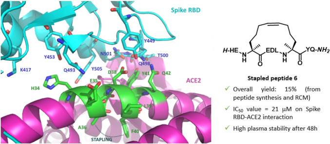
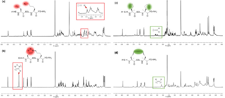
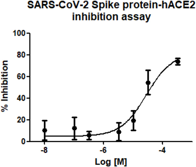
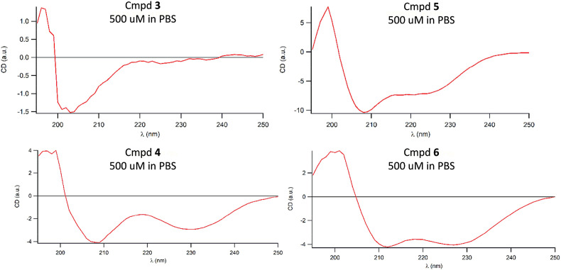
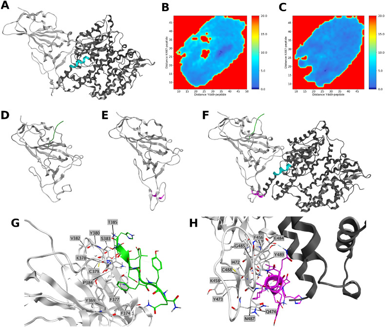
The design and synthesis of a series of peptide derivatives based on a short ACE2 α-helix 1 epitope and subsequent [i - i+4] stapling of the secondary structure resulted in the identification of a 9-mer peptide capable to compete with recombinant ACE2 towards Spike RBD in the micromolar range. Specifically, SARS-CoV-2 Spike inhibitor screening based on colorimetric ELISA assay and structural studies by circular dichroism showed the ring-closing metathesis cyclization being capable to stabilize the helical structure of the 9-mer 34HEAEDLFYQ42 epitope better than the triazole stapling via click chemistry. MD simulations showed the stapled peptide being able not only to bind the Spike RBD, sterically interfering with ACE2, but also showing higher affinity to the target as compared to parent epitope.

Keywords: COVID-19; Infectious diseases; Molecular modeling; Peptidomimetics; Protein-protein interaction; Ring-closing metathesis.


Conflict of interest statement

Declaration of competing interest The authors declare that they have no known competing financial interests or personal relationships that could have appeared to influence the work reported in this paper.

Figures

Image 1
Graphical abstract
Fig. 1
Fig. 1
Left: trimeric Spike protein (cyan) complexed to two ACE2 fragments (purple, PDB: 7A97); right: close-up of RBD (cyan)-ACE2 (purple) interaction and highlight of the selected ACE2 α-helix 1 epitope (green, PDB: 6M0J).
Fig. 2
Fig. 2
Magnification of RBD (cyan)-ACE2 (purple) interaction, highlighting the selected ACE2 α-helix 1 epitope (green, PDB: 6M0J), the key interacting amino acids and the selected amino acids for the [i-i+4] stapling region.
Fig. 3
Fig. 3
Left: plots of 1H NMR spectra of (a) acyclic peptide 3 and (b) stapled peptide 4 with box highlighting the key signals of alkyne and triazole protons. Right: plots of 1H NMR spectra of (c) acyclic peptide 5 and (b) stapled peptide 6 with box highlighting the key signals of terminal alkenes and internal double bond protons. Spectra were recorded at 400 MHz in D2O.
Fig. 4
Fig. 4
Inhibition curve of 6 on Spike RBD-ACE2 interaction.
Fig. 5
Fig. 5
Circular dichroism spectra of triazole-stapled peptide 4 and RCM-stapled peptide 6 along with linear precursors 3 and 5, respectively, at 500 μM in PBS. Spectra were recorded between 190 and 250 nm.
Fig. 6
Fig. 6
The plasma stability of 1 and 6 at 37 °C recording peptide degradation over time using LC-MS/MS.
Fig. 7
Fig. 7
A) 3D structure of SpikeRBD:ACE2 complex (PDB entry: 6M0J). Spike RBD is represented in light grey ribbon, ACE2 is rendered in dark grey ribbon. Sequence of the protein consisting of WT peptide is highlighted in cyan; B–C) PMFs obtained from distances of CA atoms of residues Y449 and K487 to CA atoms of B) 1 and C) 6. PMF is shown in color bar (0.0–20.0 kcal/mol range) from dark blue to red; D) Lowest energy conformation obtained from the simulation of SpikeRBD:1 complex. SpikeRBD is represented in light grey, WT is rendered in green; E) Lowest energy conformation obtained from the simulation of SpikeRBD:6 complex. SpikeRBD is represented in light grey, 6 is rendered in purple; F) Superposition of 1 and 6 to SpikeRBD:ACE2 complex. Spike RBD is represented in light grey ribbon ACE2 is rendered in dark grey ribbon. Sequence of the protein consisting of 1 is highlighted in cyan, 1 is rendered in green, 6 is rendered in purple; G) Site view of the interactions of SpikeRBD:1 PMF = 0 geometry. Spike RBD amino acids within 4.5 Å of WT are represented in grey sticks. 1 residues are rendered in green. Spike RBD is represented in light grey ribbon; H) Site view of the interactions of SpikeRBD:6 PMF = 0 geometry. ACE2 is rendered in dark grey ribbon Spike RBD amino acids within 4.5 Å of 6 are represented in purple sticks. Peptide 6 residues are rendered in purple. Spike RBD is represented in light grey ribbon.

References

    1. Zhou P., Yang X.L., Wang X.G., Hu B., Zhang L., Zhang W., Si H.R., Zhu Y., Li B., Huang C.L., et al. A pneumonia outbreak associated with a new coronavirus of probable bat origin. Nature. 2020;579:270–273. - PMC - PubMed
    1. Wu F., Zhao S., Yu B., Chen Y.M., Wang W., Song Z.G., Hu Y., Tao Z.W., Tian J.H., Pei Y.Y., et al. A new coronavirus associated with human respiratory disease in China. Nature. 2020;579:265–269. - PMC - PubMed
    1. WHO, Coronavirus disease (COVID-19) pandemic. https://www.who.int/emergencies/diseases/novel-coronavirus-2019
    1. Worldometer, Cases Coronavirus. https://www.worldometers.info/coronavirus/coronavirus-cases/#daily-cases
    1. Callaway E. Beyond Omicron: what's next for COVID's viral evolution. Nature. 2021;600:204–207. - PubMed