

Effective Mechanical Thrombectomy for Posterior Circulation Ischemia Using Magnetic Resonance Imaging-based Arterial Structures
- PMID: 36682792
- PMCID: PMC10072885
- DOI: 10.2176/jns-nmc.2022-0246
Effective Mechanical Thrombectomy for Posterior Circulation Ischemia Using Magnetic Resonance Imaging-based Arterial Structures
Abstract
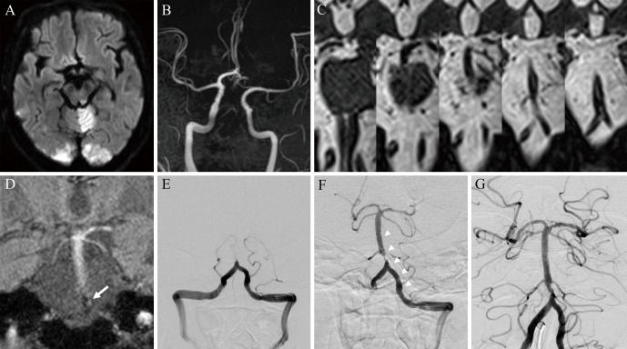
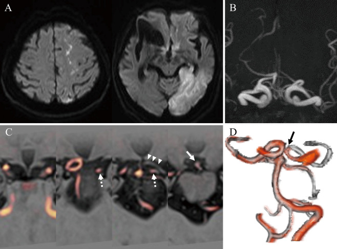
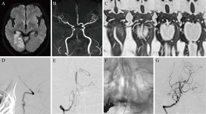
To improve the success of mechanical thrombectomy, three-dimensional turbo spin-echo (3D-TSE) sequences on T2WI can be employed to estimate the vascular structure of the posterior circulation. In addition to the short imaging time of 3D-TSE T2WI (33 sec), it can visualize the outer diameter of the main cerebral artery, including the occluded vessels. However, to date, the efficacy of mechanical thrombectomy in the posterior circulation remains unclear, and safer and more efficient mechanical thrombectomy procedures are required. Assessment of the anatomical variations in the posterior circulation using 3D-TSE T2WI is valuable for access decisions, device selection, and safe device guidance and retrieval techniques to the target vessel. Herein, we present representative cases of basilar artery and posterior cerebral artery occlusions in our institute and describe the utility of preoperative 3D-TSE T2WI in these patients.
Keywords: 3D turbo spin-echo magnetic resonance imaging; mechanical thrombectomy; posterior circulation ischemia.
Conflict of interest statement
All authors declare no conflicts of interest associated with this manuscript.
Figures
Similar articles
-
Evaluation of Occluded Distal Vessels with Variable Flip-Angle 3-Dimensional Turbo Spin-Echo Magnetic Resonance Imaging Before Acute Mechanical Thrombectomy.World Neurosurg. 2022 Nov;167:9-16. doi: 10.1016/j.wneu.2022.08.085. Epub 2022 Aug 24. World Neurosurg. 2022. PMID: 36030009
-
3D Turbo Spin-echo MRI-based Mechanical Thrombectomy at Middle Cerebral Artery Bifurcations.Neurol Med Chir (Tokyo). 2022 Mar 15;62(3):149-155. doi: 10.2176/nmc.tn.2021-0179. Epub 2021 Dec 8. Neurol Med Chir (Tokyo). 2022. PMID: 34880196 Free PMC article.
-
Three-dimensional vessel wall MRI to characterize thrombus prior to endovascular thrombectomy for large vessel occlusion stroke.J Neuroimaging. 2022 Nov;32(6):1070-1074. doi: 10.1111/jon.13050. Epub 2022 Sep 18. J Neuroimaging. 2022. PMID: 36117145
-
A novel route of revascularization in basilar artery occlusion and review of the literature.J Neurointerv Surg. 2016 Jul;8(7):e25. doi: 10.1136/neurintsurg-2015-011723.rep. Epub 2015 Jun 10. J Neurointerv Surg. 2016. PMID: 26063797 Review.
-
[Endovascular treatment of acute basilar artery occlusions].Nervenarzt. 2021 Aug;92(8):752-761. doi: 10.1007/s00115-021-01123-y. Epub 2021 May 3. Nervenarzt. 2021. PMID: 33938960 Review. German.
References
-
- Markus HS, van der Worp HB, Rothwell PM: Posterior circulation ischaemic stroke and transient ischaemic attack: Diagnosis, investigation, and secondary prevention. Lancet Neurol 12: 989-998, 2013 - PubMed
-
- Mattle HP, Arnold M, Lindsberg PJ, Schonewille WJ, Schroth G: Basilar artery occlusion. Lancet Neurol 10: 1002-1014, 2011 - PubMed
-
- Powers WJ, Rabinstein AA, Ackerson T, et al. : Guidelines for the early management of patients with acute ischemic stroke: 2019 update to the 2018 guidelines for the early management of acute ischemic stroke: A guideline for healthcare professionals from the American Heart Association/American Stroke Association. Stroke 50: 344-418, 2019 - PubMed
-
- Liu X, Dai Q, Ye R, et al. : Endovascular treatment versus standard medical treatment for vertebrobasilar artery occlusion (BEST): an open-label, randomised controlled trial. Lancet Neurol 19: 115-122, 2020 - PubMed
-
- Langezaal LCM, van der Hoeven EJRJ, Mont'Alverne FJA, et al. : Endovascular therapy for stroke due to basilar-artery occlusion. N Engl J Med 384: 1910-1920, 2021 - PubMed