

Rapid Diagnostic Tests for the Detection of the Four Dengue Virus Serotypes in Clinically Relevant Matrices
- PMID: 36682882
- PMCID: PMC9927141
- DOI: 10.1128/spectrum.02796-22
Rapid Diagnostic Tests for the Detection of the Four Dengue Virus Serotypes in Clinically Relevant Matrices
Abstract
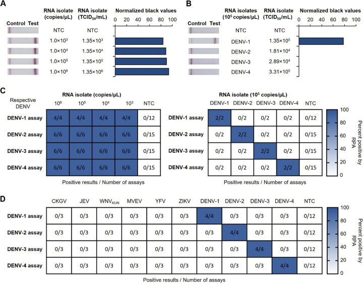
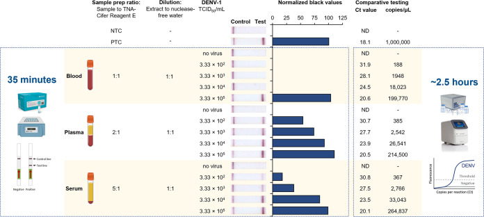
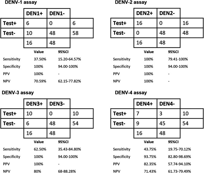
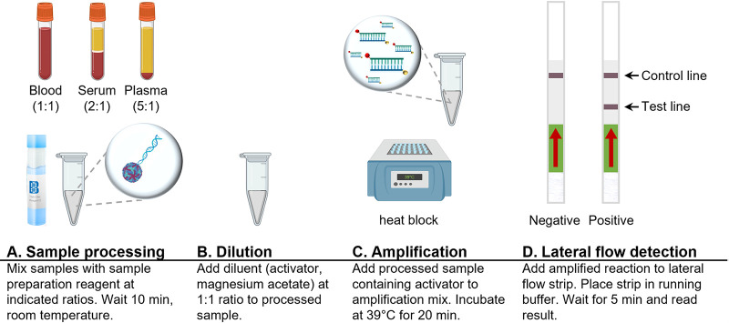
The efficient and accurate diagnosis of dengue, a major mosquito-borne disease, is of primary importance for clinical care, surveillance, and outbreak control. The identification of specific dengue virus serotype 1 (DENV-1) to DENV-4 can help in understanding the transmission dynamics and spread of dengue disease. The four rapid low-resource serotype-specific dengue tests use a simple sample preparation reagent followed by reverse transcription-isothermal recombinase polymerase amplification (RT-RPA) combined with lateral flow detection (LFD) technology. Results are obtained directly from clinical sample matrices in 35 min, requiring only a heating block and pipettes for liquid handling. In addition, we demonstrate that the rapid sample preparation step inactivates DENV, improving laboratory safety. Human plasma and serum were spiked with DENV, and DENV was detected with analytical sensitivities of 333 to 22,500 median tissue culture infectious doses (TCID50)/mL. The analytical sensitivities in blood were 94,000 to 333,000 TCID50/mL. Analytical specificity testing confirmed that each test could detect multiple serotype-specific strains but did not respond to strains of other serotypes, closely related flaviviruses, or chikungunya virus. Clinical testing on 80 human serum samples demonstrated test specificities of between 94 and 100%, with a DENV-2 test sensitivity of 100%, detecting down to 0.004 PFU/μL, similar to the sensitivity of the PCR test; the other DENV tests detected down to 0.03 to 10.9 PFU/μL. Collectively, our data suggest that some of our rapid dengue serotyping tests provide a potential alternative to conventional labor-intensive RT-quantitative PCR (RT-qPCR) detection, which requires expensive thermal cycling instrumentation, technical expertise, and prolonged testing times. Our tests provide performance and speed without compromising specificity in human plasma and serum and could become promising tools for the detection of high DENV loads in resource-limited settings. IMPORTANCE The efficient and accurate diagnosis of dengue, a major mosquito-borne disease, is of primary importance for clinical care, surveillance, and outbreak control. This study describes the evaluation of four rapid low-resource serotype-specific dengue tests for the detection of specific DENV serotypes in clinical sample matrices. The tests use a simple sample preparation reagent followed by reverse transcription-isothermal recombinase polymerase amplification (RT-RPA) combined with lateral flow detection (LFD) technology. These tests have several advantages compared to RT-qPCR detection, such as a simple workflow, rapid sample processing and turnaround times (35 min from sample preparation to detection), minimal equipment needs, and improved laboratory safety through the inactivation of the virus during the sample preparation step. The low-resource formats of these rapid dengue serotyping tests have the potential to support effective dengue disease surveillance and enhance the diagnostic testing capacity in resource-limited countries with both endemic dengue and intense coronavirus disease 2019 (COVID-19) transmission.
Keywords: NS5 gene; dengue virus; isothermal amplification; lateral flow detection; rapid molecular assays; rapid sample preparation; recombinase polymerase amplification.
Conflict of interest statement
The authors declare a conflict of interest. Pollak N.M. is a funded post-doctoral research scientist for DMTC Ltd, Australia. Macdonald J. is a Project Leader for DMTC Ltd, Australia. Macdonald J. is a co-founder, shareholder, director and employee of BioCifer Pty Ltd, who co-funded the study and licensed the technology. All other authors declare no competing interest.
Figures

Similar articles
-
Development of a rapid point-of-care dengue virus type 2 infection diagnostic assay using recombinase polymerase amplification and lateral flow device.Front Cell Infect Microbiol. 2025 May 14;15:1578549. doi: 10.3389/fcimb.2025.1578549. eCollection 2025. Front Cell Infect Microbiol. 2025. PMID: 40438242 Free PMC article.
-
Rapid and visual detection of dengue virus using recombinase polymerase amplification method combined with lateral flow dipstick.Mol Cell Probes. 2019 Aug;46:101413. doi: 10.1016/j.mcp.2019.06.003. Epub 2019 Jun 14. Mol Cell Probes. 2019. PMID: 31202830
-
Validation of the Pockit Dengue Virus Reagent Set for Rapid Detection of Dengue Virus in Human Serum on a Field-Deployable PCR System.J Clin Microbiol. 2018 Apr 25;56(5):e01865-17. doi: 10.1128/JCM.01865-17. Print 2018 May. J Clin Microbiol. 2018. PMID: 29436418 Free PMC article.
-
Analytical and diagnostic performance characteristics of reverse-transcriptase loop-mediated isothermal amplification assays for dengue virus serotypes 1-4: A scoping review to inform potential use in portable molecular diagnostic devices.PLOS Glob Public Health. 2023 Aug 8;3(8):e0002169. doi: 10.1371/journal.pgph.0002169. eCollection 2023. PLOS Glob Public Health. 2023. PMID: 37552632 Free PMC article.
-
Portable Point-of-Care Diagnosis Platforms and Emerging Predictive Biomarkers for Rapid Detection of Severe Dengue Viral Infection.ACS Sens. 2025 May 23;10(5):3302-3316. doi: 10.1021/acssensors.5c00263. Epub 2025 Mar 31. ACS Sens. 2025. PMID: 40165016 Free PMC article. Review.
Cited by
-
Evaluation of three rapid low-resource molecular tests for Nipah virus.Front Microbiol. 2023 Feb 9;13:1101914. doi: 10.3389/fmicb.2022.1101914. eCollection 2022. Front Microbiol. 2023. PMID: 36845977 Free PMC article.
-
Transfusion-Transmitted Disorders 2023 with Special Attention to Bone Marrow Transplant Patients.Pathogens. 2023 Jul 1;12(7):901. doi: 10.3390/pathogens12070901. Pathogens. 2023. PMID: 37513748 Free PMC article. Review.
-
The use of rapid diagnostic tests for chronic Chagas disease: An expert meeting report.PLoS Negl Trop Dis. 2024 Aug 8;18(8):e0012340. doi: 10.1371/journal.pntd.0012340. eCollection 2024 Aug. PLoS Negl Trop Dis. 2024. PMID: 39116064 Free PMC article. Review.
-
Rapid and sensitive detection of chikungunya virus using one-tube, reverse transcription, semi-nested multi-enzyme isothermal rapid amplification, and lateral flow dipstick assays.J Clin Microbiol. 2024 Sep 11;62(9):e0038324. doi: 10.1128/jcm.00383-24. Epub 2024 Aug 14. J Clin Microbiol. 2024. PMID: 39140738 Free PMC article.
-
Rapid, sensitive, and specific, low-resource molecular detection of Hendra virus.One Health. 2023 Feb 10;16:100504. doi: 10.1016/j.onehlt.2023.100504. eCollection 2023 Jun. One Health. 2023. PMID: 37363221 Free PMC article.
References
-
- World Health Organization. 2019. Dengue increase likely during rainy season: WHO warns. World Health Organization, Geneva, Switzerland. https://www.who.int/westernpacific/news/detail/11-06-2019-dengue-increas.... Accessed 16 May 2022.
-
- Messina JP, Brady OJ, Golding N, Kraemer MUG, Wint GRW, Ray SE, Pigott DM, Shearer FM, Johnson K, Earl L, Marczak LB, Shirude S, Davis Weaver N, Gilbert M, Velayudhan R, Jones P, Jaenisch T, Scott TW, Reiner RC, Jr, Hay SI. 2019. The current and future global distribution and population at risk of dengue. Nat Microbiol 4:1508–1515. doi:10.1038/s41564-019-0476-8. - DOI - PMC - PubMed
Publication types
MeSH terms
Substances
LinkOut - more resources
Full Text Sources
Medical
Research Materials
