

Ventriculus terminalis cyst in an infant: a case report
- PMID: 36683067
- PMCID: PMC9869499
- DOI: 10.1186/s13256-023-03759-7
Ventriculus terminalis cyst in an infant: a case report
Abstract
Background: Filar cysts are frequently found on neonatal ultrasound and are physiologically involuting structures with natural resolution. Hence, there has been no previous histologic correlation. Ventriculus terminalis is a focal central canal dilation in the conus medullaris and usually not clinically significant. Extra-axial cyst at the conus-filum junction connected to ventriculus terminalis is extremely rare, especially when associated with tethered lipomatous filum terminale and with progressive cyst enlargement.
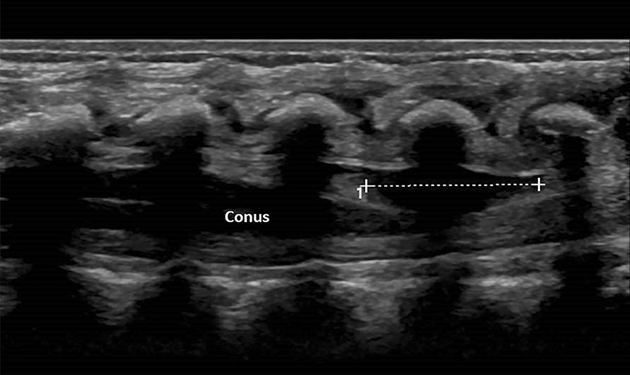
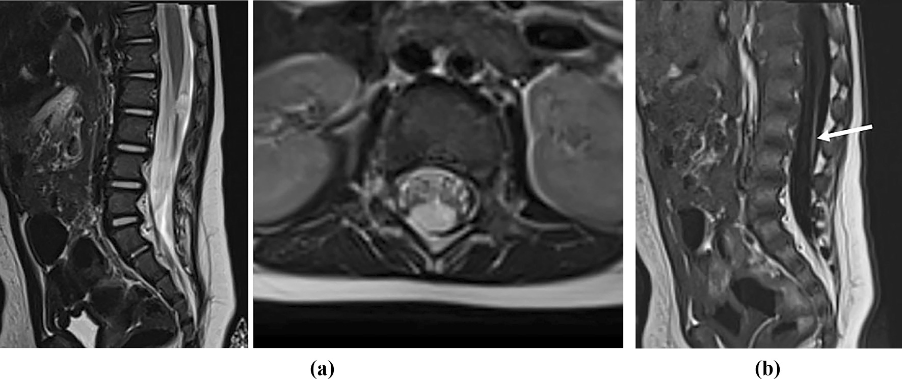
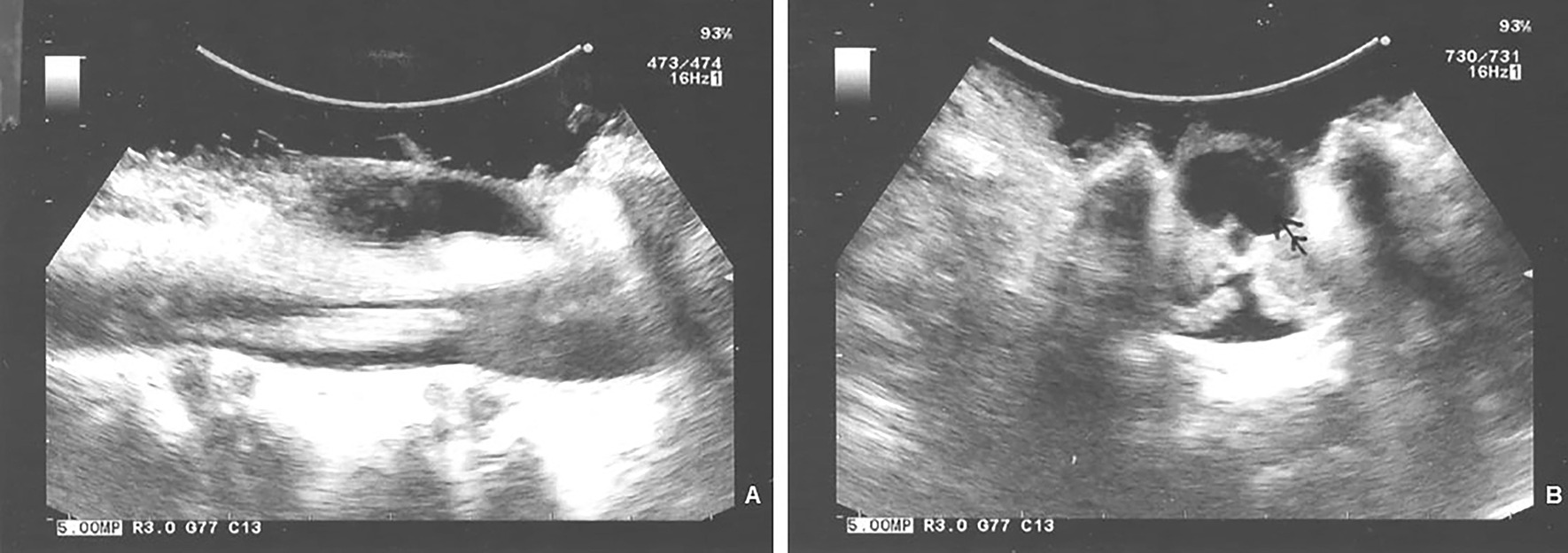
Case presentation: A Caucasian female neonate with abnormal gluteal cleft had ventriculus terminalis cyst with an extra-axial cyst at the conus-filar junction and taut lipomatous filum on ultrasound examination and magnetic resonance imaging. This persisted at 6-month follow up imaging. In light of the nonresolving extra-axial mass and thick taut lipomatous filum, the child underwent L1-L3 osteoplastic laminectomies. The extra-axial cyst expanded after bony decompression and furthermore on dural opening; visualized on ultrasound. It communicated with the central canal and was documented with intraoperative photomicrographs. It was excised and filum sectioned. Histological immunostaining of the cyst wall showed neuroglial and axonal elements. The child did well without deficits at 4-year follow up with normal urodynamics.
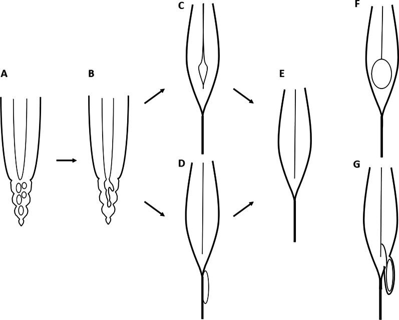
Conclusion: Progression dilation of ventriculus terminalis and extra-axial conofilar cyst with tethered lipomatous filum will likely progress to clinical significance and require surgical intervention. The embryologic basis for this pathology is discussed, with literature review.
Keywords: Case report; Extra-axial cauda equina cyst; Filar cyst; Lipomatous filum; Tethered cord; Ventriculus terminalis.
© 2023. The Author(s).
Conflict of interest statement
The authors report no conflicts of interest concerning the material or methods used in this study or the findings specified in this paper.
Figures

References
-
- Kernohan JW. The ventriculus terminalis: its growth and development. J Comp Neurol. 1924;38:107–125. doi: 10.1002/cne.900380106. - DOI
Publication types
MeSH terms
LinkOut - more resources
Full Text Sources
