Review
. 2023 Apr;70(4):e30219.
doi: 10.1002/pbc.30219. Epub 2023 Jan 22.

Kaposiform lymphangiomatosis: Diagnosis, pathogenesis, and treatment

Affiliations
Review

Kaposiform lymphangiomatosis: Diagnosis, pathogenesis, and treatment

C Griffin McDaniel et al. Pediatr Blood Cancer. 2023 Apr.

Abstract

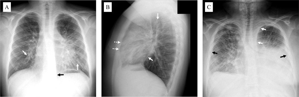
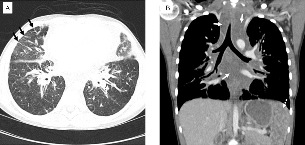
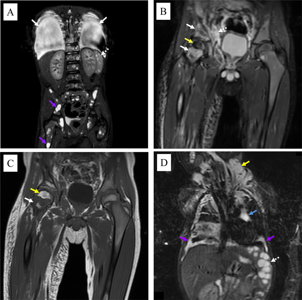
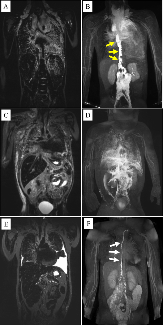
Kaposiform lymphangiomatosis (KLA) is a life-threatening rare disease that can cause substantial morbidity, mortality, and social burdens for patients and their families. Diagnosis often occurs long after initial symptoms, and there are few centers in the world with the expertise to diagnose and care for patients with the disease. KLA is a lymphatic anomaly and significant advancements have been made in understanding its pathogenesis and etiology since its first description in 2014. This review provides multidisciplinary, comprehensive, and state-of-the-art information on KLA patient presentation, diagnostic imaging, pathology, organ involvement, genetics, and pathogenesis. Finally, we describe current therapeutic approaches, important areas for research, and challenges faced by patients and their families. Further insights into the pathogenesis of KLA may advance our understanding of other vascular anomalies given that similar signaling pathways may be involved.

Keywords: kaposiform lymphangiomatosis; lymphatic anomalies; rare diseases.


Conflict of interest statement

Conflict of interest disclosure: DMA is a consultant and steering committee member for Novartis and a consultant for Alderis and Nobias. AMH provides clinical research support for Merck (ongoing), Novartis (ongoing), Venthera (within the past year), and Palvella (within the past year); is a steering committee member for Novartis; and is a consultant for Novartis, Ideaya, and Aytu. ACM is an author/consultant/editor for Elsevier, Inc for royalties and fees.

Figures

Figure 1.
Figure 1.
Chest radiography of patients with kaposiform lymphangiomatosis. A) Upright posteroanterior view in a 12-year-old male shows thickening of the peribronchovascular interstitium (solid white arrows). Widening of the left paraspinal stripe inferiorly (solid black arrow) is due to mediastinal infiltration by the disease. B) Upright lateral chest view in the same patient shows thickening of the peribronchovascular interstitium (solid white arrows) with visualization of peripheral interlobular septal thickening anteriorly (dotted white arrows). C) Supine frontal chest view in a 25-year-old male shows generalized interstitial thickening with left greater than right pleural effusions (solid black arrows). There is also mediastinal widening due to infiltration by abnormal lymphatic channels/fluid (solid white arrows).
Figure 2.
Figure 2.
Computed tomography images of patient with kaposiform lymphangiomatosis. A) Axial contrast-enhanced chest image in lung windows in an 11-year-old female shows variable degrees of interstitial thickening, visible along the bronchial walls centrally (white arrows) and secondary pulmonary lobules peripherally (black arrows). B) Coronal contrast-enhanced chest image in the same patient shows expansion of the mediastinum by poorly defined fluid attenuation tissue (solid white arrows), corresponding to infiltration by disease. Numerous lesions are also noted in the spleen (dotted white arrow).
Figure 3.
Figure 3.
Coronal magnetic resonance imaging of patients with kaposiform lymphangiomatosis. A) Short-T1 inversion recovery (STIR) image of the body in a 5-year-old male shows large pleural effusions (solid white arrows), multiple small cystic-appearing splenic lesions (dotted white arrows), and numerous cystic-appearing lesions throughout the skeleton (purple arrows). B) STIR image in the same patient shows abnormal confluent foci of increased signal intensity throughout the right pelvis and femoral neck (solid white arrows). There is also abnormally decreased signal intensity throughout the right femoral head (yellow arrow). A more normal pattern of red marrow is seen in most of the left pelvis and femur. Note also the abnormal poorly defined lymphatic tissue in the pelvic soft tissues (dotted white arrow). C) T1 image in the same patient shows intermixed regions of increased signal intensity (yellow arrow) due to increased fat deposition as well as regions of decreased signal intensity (solid white arrow) that are more typical of abnormal lymphatics. D) STIR image in a 10-month-old male shows cystic-appearing splenic lesions (dotted white arrow). Bilateral pleural disease (purple arrows) is noted, as well as mediastinal (blue arrow) and neck (yellow arrow) lymphatic malformations.
Figure 4.
Figure 4.
Coronal T2-weighted magnetic resonance imaging (MRI) and magnetic resonance lymphangiography (MRL) of patients with kaposiform lymphangiomatosis. A) T2-weighted MRI of a 10 year-old male shows significant mediastinal thickening with pulmonary edema and bilateral supraclavicular edema. B) The corresponding MRL shows a significantly dilated and tortuous thoracic duct (yellow arrows) that terminates in the mediastinum with significant perfusion of the mediastinal and pulmonary lymphatics on MRL. C) T2-weighted MRI of a 4 year-old male shows mediastinal thickening with pulmonary edema, dermal edema, and ascites. D) The corresponding MRL shows no clear thoracic duct but several abnormal lymphatic channels coursing into the thorax leading to mediastinal and pulmonary lymphatic perfusion. E) T2-weighted MRI of a 12 year-old female shows mediastinal thickening with bilateral pleural effusions. F) The corresponding MRL shows a dilated and mildly tortuous thoracic duct (white arrows), but with limited mediastinal or pulmonary lymphatic perfusion.
Figure 5.
Figure 5.
Histopathology of kaposiform lymphangiomatosis lesions. A) Lung with marked dilated lymphatic channels in pleura and interlobular septa. B) Lung with cluster of spindled cells and interspersed erythrocytes. C) Single, dilated lymphatic channel in reticular dermis and small cellular cluster in middle of the field. D) Dermal cellular cluster of hemosiderotic spindled cells with interspersed erythrocytes. E) Cytoplasmic immunopositivity for D2–40 in spindled cell cluster and adjacent dilated lymphatic vessels in lung. F) Nuclear immunopositivity for PROX1 in splenic sinusoidal lining cells. Dermal spindled cell clusters with cytoplasmic immunopositivity for D2–40 (G) and LYVE1 (H), and nuclear immunopositivity for PROX1 (I). J) Spleen with markedly expanded cords of Billroth and dilated sinusoids. K) Splenic hyperplastic sinusoidal cells, some with phagocytosed red blood cells and cytoplasmic eosinophilic globules.

References

    1. Croteau SE, Kozakewich HP, Perez-Atayde AR, et al. Kaposiform lymphangiomatosis: a distinct aggressive lymphatic anomaly. J Pediatr. 2014;164(2):383–388. - PMC - PubMed
    1. Perez-Atayde AR, Debelenko L, Al-Ibraheemi A, et al. Kaposiform Lymphangiomatosis: Pathologic Aspects in 43 Patients. Am J Surg Pathol. 2022;46(7):963–976. - PubMed
    1. Adams DM, Trenor CC, Hammill AM, et al. Efficacy and Safety of Sirolimus in the Treatment of Complicated Vascular Anomalies. Pediatrics. 2016;137(2):e20153257. - PMC - PubMed
    1. Fernandes VM, Fargo JH, Saini S, et al. Kaposiform lymphangiomatosis: unifying features of a heterogeneous disorder. Pediatr Blood Cancer. 2015;62(5):901–904. - PubMed
    1. Wang Z, Li K, Yao W, et al. Successful treatment of kaposiform lymphangiomatosis with sirolimus. Pediatric blood & cancer. 2015;62(7):1291–1293. - PubMed