. 2023 Apr;238(2):654-672.
doi: 10.1111/nph.18756. Epub 2023 Feb 18.

DELLA proteins regulate spore germination and reproductive development in Physcomitrium patens

Affiliations

DELLA proteins regulate spore germination and reproductive development in Physcomitrium patens

Alexandros Phokas et al. New Phytol. 2023 Apr.

Abstract

Proteins of the DELLA family integrate environmental signals to regulate growth and development throughout the plant kingdom. Plants expressing non-degradable DELLA proteins underpinned the development of high-yielding 'Green Revolution' dwarf crop varieties in the 1960s. In vascular plants, DELLAs are regulated by gibberellins, diterpenoid plant hormones. How DELLA protein function has changed during land plant evolution is not fully understood. We have examined the function and interactions of DELLA proteins in the moss Physcomitrium (Physcomitrella) patens, in the sister group of vascular plants (Bryophytes). PpDELLAs do not undergo the same regulation as flowering plant DELLAs. PpDELLAs are not degraded by diterpenes, do not interact with GID1 gibberellin receptor proteins and do not participate in responses to abiotic stress. PpDELLAs do share a function with vascular plant DELLAs during reproductive development. PpDELLAs also regulate spore germination. PpDELLAs interact with moss-specific photoreceptors although a function for PpDELLAs in light responses was not detected. PpDELLAs likely act as 'hubs' for transcriptional regulation similarly to their homologues across the plant kingdom. Taken together, these data demonstrate that PpDELLA proteins share some biological functions with DELLAs in flowering plants, but other DELLA functions and regulation evolved independently in both plant lineages.

Keywords: Physcomitrium patens; DELLA proteins; diterpenes; light receptors; reproduction; spores; transcriptional regulation.


Conflict of interest statement

None declared.

Figures

Fig. 1
Fig. 1
Physcomitrium patens PpDELLAs form a monophyletic group with other bryophyte DELLAs and have a divergent DELLA‐LEQLE‐VNHYP domain compared to vascular plants with a conserved LHR1 region in the GRAS domain. (a) Maximum likelihood phylogenetic tree generated using the peptide sequences of selected DELLA homologues from bryophytes, lycophytes, ferns, gymnosperms and angiosperms. The monophyletic bryophyte group is shown in red. Scale bar, 0.1 substitutions per amino acid site. Numbers indicate bootstrap values at the nodes. (b) Alignment of the N‐terminal DELLA‐LEQLE‐VHNYP domain that is necessary for the interaction with GID1 receptors in angiosperms. (c) Alignment of the LHR1 region of the GRAS domain demonstrates similarity with GRAS domains from vascular plants and other bryophytes. In (b, c) black shading indicates that at least 50% of the amino acids in a particular column are identical. Amino acids that are similar to the column‐consensus peptide are shaded grey. The peptide sequences used in this figure are as follows: Arabidopsis thaliana, Medicago truncatula, Solanum lycopersicum, Hordeum vulgare, Triticum aestivum, Zea mays, Oryza sativa, Amborella trichopoda (angiosperms), Pinus tabuliformis (gymnosperm), Ceratopteris richardii (fern), Selaginella kraussiana, Selaginella moellendorfii (lycophytes), Encalypta streptocarpa, Timmia austriaca, Hedwigia ciliata, Schwetschkeopsis fabronia, Physcomitrium patens, Sphagnum fallax (mosses), Marchantia polymorpha (liverwort), Anthoceros agrestis and Anthoceros punctatus (hornworts).
Fig. 2
Fig. 2
Physcomitrium patens PpDELLA proteins are not degraded by diterpenes. (a) Top panels: GFP‐AtRGA is degraded in 7‐d‐old Arabidopsis (pRGA::GFP‐AtRGA) roots following 2‐h incubation with 10 μM gibberellin A3 (GA3). Middle panels: PpDELLAa‐GFP is not degraded in 7‐d‐old P. patens protonemata following 2‐h incubation with either 10 μM GA3 or 10 μM GA9 methyl ester (GA9‐ME). White arrowheads: nuclear PpDELLAa‐GFP. Bottom panels: PpDELLAb‐GFP is not degraded in 7‐d‐old P. patens protonemata following 2‐h incubation with either 10 μM GA3 or 10 μM GA9‐ME. Scale bars, 50 μM. (b) GFP‐AtRGA is degraded in 7‐d‐old Arabidopsis (pRGA::GFP‐AtRGA) roots following 2‐h incubation with 10 μM GA3 while PpDELLAa‐GFP and PpDELLAb‐GFP are not degraded in 7‐d‐old P. patens protonema tissue following 2‐h incubation with either 10 μM GA3 or 10 μM GA9‐ME. CBB, Coomassie Brilliant Blue staining.
Fig. 3
Fig. 3
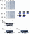
Physcomitrium patens PpDELLAs do not interact with a moss GID1‐like protein or with an Arabidopsis GID1 gibberellin receptor and an AtDELLA protein does not interact with a putative moss GID1‐like protein. (a) Upper panel: in a yeast (Saccharomyces cerevisiae) two‐hybrid assay, Arabidopsis AtRGA and AtGID1c interact with one another only in the presence of the gibberellin GA3, but not GA9 methyl ester (GA9‐ME) or ent‐kaurenoic acid (ent‐KA) or a solvent control (Mock). In the same system, PpDELLAa and PpDELLAb do not interact with PpGLP1 including in the presence of diterpenes, AtRGA does not interact with PpGLP1 and PpDELLAa/PpDELLAb do not interact with AtGID1c under any conditions. All DELLA proteins were cloned into the yeast vector pGADT7 while GID1 proteins were cloned into the pGBKT7 vector. Lower panel: no autoactivation is seen when each construct is transformed into yeast alongside the corresponding empty vector. In both panels, –LW is growth in the absence of leucine and tryptophan (to test for the presence of the plasmids) while –LWAH is growth in the absence of leucine, tryptophan, adenine and histidine which tests additionally for the protein–protein interaction. (b) Western blot of yeast cell extracts confirming that HA‐tagged PpDELLAs and AtRGA and MYC‐tagged PpGLP1 and AtGID1 are expressed in yeast: DELLA proteins detected using anti‐HA and receptor proteins detected using anti‐MYC. CBB, Coomassie Brilliant Blue staining. (c) Coimmunoprecipitation from an in vitro cell free system using α‐MYC‐coupled beads. HA‐AtRGA and MYC‐AtGID1c interacted in a GA3‐dependent manner (arrowhead, faint 67kDa band; *, antibody heavy chain), whereas HA‐PpDELLAs and MYC‐PpGLP1 did not interact in the presence or absence of GA9‐ME (*, antibody heavy chain).
Fig. 4
Fig. 4
Physcomitrium patens Ppdella mutants germinate faster than wild type but are sensitive to application of diterpenes. (a) Ppdellaa mutant spores germinate faster than wild type (WT). A Z‐test indicates significant differences between Ppdellaa and WT on days 4, 7, 8, 9 and 11. (b) Ppdellab mutant spores germinate faster than WT. A Z‐test indicates significant differences between Ppdellaa and WT on days 4, 7, 8, 9 and 11. (c) Ppdellaab mutant spores germinate faster than WT. A Z‐test indicates significant differences between Ppdellaa and WT on days 3, 4, 7, 8, 9, 11 and 16. (d) Treatment with 5 μM GA9‐ME increases spore germination rate of Ppdellaa and WT to a similar extent. A Kruskal‐Wallis test indicates significant differences between Ppdellaa + GA9‐ME and WT + methanol on days 4 and 5 (P < 0.01), between Ppdellaa + GA9‐ME and WT + GA9‐ME on day 4 (P < 0.05), between Ppdellaa + methanol and WT + methanol on day 5 (P < 0.05), between WT + GA9‐ME and WT + methanol (P < 0.05) on day 7, and between Ppdellaa + methanol and WT + GA9‐ME on day 7 (P < 0.05). (e) Treatment with GA9‐ME increases spore germination rate of Ppdellab and WT to a similar extent. A Kruskal–Wallis test indicates significant differences between Ppdellab + GA9‐ME and WT + methanol on days 4, 5 and 7 (P < 0.05). (f) Wild type and Ppdellaab spores treated with 10 μM ABA show a similar extent of germination suppression. A Kruskal‐Wallis test indicates significant differences between Ppdellaab + ABA and Ppdellaab + methanol on days 3, 4, 5 and 7, and between Ppdellaab + methanol and WT + ABA on 3, 4, 5 and 7 (P < 0.05). All germination assays are representative of three or more biological repeats. Error bars, ±SE.
Fig. 5
Fig. 5
Physcomitrium patens Ppdellaab mutants show defective sporophyte development. (a) Ppdellaab mutants (right) make fewer sporophytes than wild type (WT, left) plants under conditions that induce sex organ development, fertilisation and sporophyte development (7 wk at 15°C with 8 h light). Scale bar, 2 mm. Representative of six biological repeats across two laboratories. (b) WT plants have higher sporophyte density than Ppdellaab plants. The number of sporophytes per cm2 plant area is shown. WT and Ppdellaab show a statistically significant difference (P = 0.0011) in sporophyte density (Mann–Whitney U test; n = 7–8). Boxes represent median and quartiles, whiskers indicate range, dots indicate individual data points and black asterisks indicate means. Representative of three biological repeats from two independent laboratories. (c) Mature gametangia and sporophyte development of WT and the mutant Ppdellaab. 21 d short day (SD): WT and Ppdellaab (mutant) both show similar amounts of immature (x, closed tip cells) and mature archegonia (*, open tips) and antheridia (yellowish color, swollen tip cell) as well as some older antheridia (brownish color). Six days after watering (daw): WT apices show embryos in stage E2 (no stomata present, Fernandez‐Pozo et al., 2020), Ppdellaab apices show several mature archegonia and mature/old antheridia. 8 daw: WT sporophyte development progressed and most sporophytes are in the ES1 stage showing developing stomata (s) and brownish color at the seta. Ppdellaab apices show old as well as some new developing gametangia. 30 daw: WT sporophytes are nearly mature, two rows of stomata (s) are present and the seta is dark brown. Ppdellaab apices show no sporophytes but multiple old and young gametangia with archegonia possessing darkened and shrunken egg cells (e). Bars, 200 μm.
Fig. 6
Fig. 6
Physcomitrium patens PpDELLA proteins interact with putative light receptors. (a) Common (743) and unique (408) proteins in anti‐GFP immunoprecipitations from samples expressing induced PpDELLAa‐GFP (blue), uninduced PpDELLAa‐GFP (yellow) or induced GFP (green). Venn diagram created with venny 2.1 (Oliveros, 2007–2015) and edited with BioRender.com. (b) Proteins immunoprecipitated with Gene Ontology (GO) biological function ‘chromophore‐protein linkage’ (GO:0018298) from PpDELLAa‐expressing plants include three photoreceptors: PHYTOCHROME5B (PpPHY5B), PHOTOTROPINA2 (PpPHOTA2) and PHOTOTROPINB1 (PpPHOTB1). (c) Yeast two‐hybrid assay between PpDELLAs fused to the GAL4 activation domain (AD) in pGADT7 and the photoreceptors: PpPHOTA2, PpPHOTB1 and PpPHY5B, fused to the GAL4 DNA‐binding (DBD) domain in pGBKT7. PpDELLAa interacted with PpPHY5B; and PpDELLAb interacted with both PpPHY5B and PpPHOTA2. No interaction between PpPHOTB1 was seen with either PpDELLAa or PpDELLAb in this system. (d) Anti‐MYC western blot showing that MYC‐tagged PpPHOTA2 (124 kDa) and PpPHY5B (126 kDa) are expressed in yeast but PpPHOTB1 (127 kDa) is not. CBB, Coomassie Brilliant Blue staining.
Fig. 7
Fig. 7
Gene Ontology (GO) term enrichment for differentially expressed genes in the Physcomitrium patens Ppdellaab mutant includes photosynthetic, metabolic and cell wall functions. (a) Genes downregulated in the Ppdellaab mutant. Colour intensity represents log fold‐change. The network represents regulatory interactions between differentially expressed genes, predicted using PlantRegMap and visualized with cytoscape. (b) Genes upregulated in the Ppdellaab mutant. Colour intensity represents log fold‐change. The network represents regulatory interactions between differentially expressed genes, predicted using PlantRegMap and visualized with cytoscape. (c) GO categories for Biological Process enriched among genes misregulated in Ppdellaab. GO term enrichment was calculated using PlantRegMap, and represented with ReviGO, based on the semantic distances between GO terms. PpDELLA‐induced biological process GO terms (from genes downregulated in the Ppdellaab mutant) include genes involved in photosynthesis and chloroplast function along with primary metabolism and responses to light and hormones. PpDELLA‐induced molecular function GO terms (from genes downregulated in the Ppdellaab mutant) are largely involved in metabolism (primary and secondary) with some genes involved in stress responses. LogSize represents the log10 (number of annotations for GO Term ID in selected species in the EBI GOA database).
Fig. 8
Fig. 8
Physcomimtrium patens PpDELLAs may act as transcriptional regulators. (a) Classes of transcription factors (TFs) most likely to be responsible for the regulation of PpDELLA‐induced genes (the TFs have putative binding sites in the promoters of genes downregulated in the Ppdellaab mutant), according to a TF‐enrichment analysis performed with PlantRegMap, visualised using cytoscape. (b) Classes of transcription factors (TFs) most likely to be responsible for the regulation of PpDELLA‐repressed genes (the TFs have putative binding sites in the promoters of genes upregulated in the Ppdellaab mutant), according to a TF‐enrichment analysis performed with PlantRegMap, visualised using cytoscape. (c) There is little overlap between the genes differentially expressed between wild type and Ppdellaab mutants, genes induced by GA9‐methyl ester (Perroud et al., 2018) and the transcription factors (DELLA‐TFs) predicted to bind to the promoters of genes differentially expressed in the Ppdellaab mutant (shown in a,b). Venn diagram produced using venny 2.1 (Oliveros, 2007–2015).

References

    1. Achard P, Cheng H, De Grauwe L, Decat J, Schoutteten H, Moritz T, Van Der Straeten D, Peng J, Harberd NP. 2006. Integration of plant responses to environmentally activated phytohormonal signals. Science 311: 91–94. - PubMed
    1. Achard P, Renou JP, Berthome R, Harberd NP, Genschik P. 2008. Plant DELLAs restrain growth and promote survival of adversity by reducing the levels of reactive oxygen species. Current Biology 18: 656–660. - PubMed
    1. Altschul SF, Gish W, Miller W, Myers EW, Lipman DJ. 1990. Basic local alignment search tool. Journal of Molecular Biology 215: 403–410. - PubMed
    1. Aya K, Hiwatashi Y, Kojima M, Sakakibara H, Ueguchi‐Tanaka M, Hasebe M, Matsuoka M. 2011. The Gibberellin perception system evolved to regulate a pre‐existing GAMYB‐mediated system during land plant evolution. Nature Communications 2: 544. - PubMed
    1. Bolger AM, Lohse M, Usadel B. 2014. Trimmomatic: a flexible trimmer for Illumina sequence data. Bioinformatics 30: 2114–2120. - PMC - PubMed

Publication types