

Pituitary Stem Cell Regulation by Zeb2 and BMP Signaling
- PMID: 36683433
- PMCID: PMC10091485
- DOI: 10.1210/endocr/bqad016
Pituitary Stem Cell Regulation by Zeb2 and BMP Signaling
Abstract
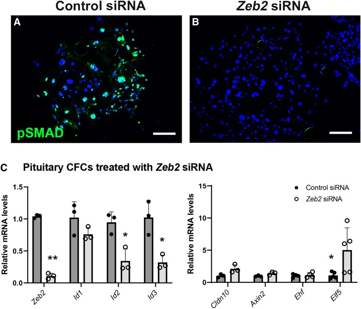
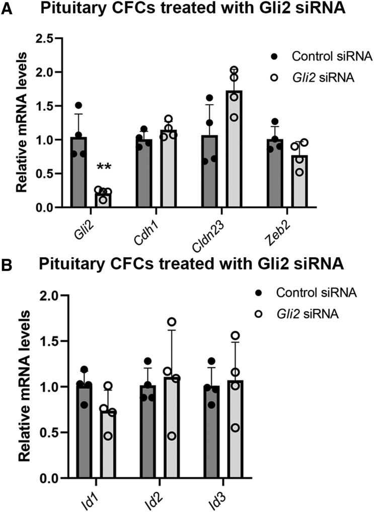
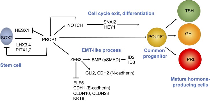
Epithelial to mesenchymal transition (EMT) is important for many developing organs, and for wound healing, fibrosis, and cancer. Pituitary stem cells undergo an EMT-like process as they migrate and initiate differentiation, but little is known about the input of signaling pathways or the genetic hierarchy of the transcriptional cascade. Prop1 mutant stem cells fail to undergo changes in cellular morphology, migration, and transition to the Pou1f1 lineage. We used Prop1 mutant mice to identify the changes in gene expression that are affiliated with EMT-like processes. BMP and TGF-β family gene expression was reduced in Prop1 mutants and Elf5, a transcription factor that characteristically suppresses EMT, had elevated expression. Genes involved in cell-cell contact such as cadherins and claudins were elevated in Prop1 mutants. To establish the genetic hierarchy of control, we manipulated gene expression in pituitary stem cell colonies. We determined that the EMT inducer, Zeb2, is necessary for robust BMP signaling and repression of Elf5. We demonstrated that inhibition of BMP signaling affects expression of target genes in the Id family, but it does not affect expression of other EMT genes. Zeb2 is necessary for expression of the SHH effector gene Gli2. However, knock down of Gli2 has little effect on the EMT-related genes, suggesting that it acts through a separate pathway. Thus, we have established the genetic hierarchy involved in the transition of pituitary stem cells to differentiation.
Keywords: ELF5; GLI2; ID gene family; Rathke's pouch; epithelial to mesenchymal-like transition; transcription factor PROP1.
© The Author(s) 2023. Published by Oxford University Press on behalf of the Endocrine Society. All rights reserved. For permissions, please e-mail: journals.permissions@oup.com.
Figures
Similar articles
-
PROP1 triggers epithelial-mesenchymal transition-like process in pituitary stem cells.Elife. 2016 Jun 28;5:e14470. doi: 10.7554/eLife.14470. Elife. 2016. PMID: 27351100 Free PMC article.
-
Premature differentiation and aberrant movement of pituitary cells lacking both Hes1 and Prop1.Dev Biol. 2009 Jan 1;325(1):151-61. doi: 10.1016/j.ydbio.2008.10.010. Epub 2008 Nov 1. Dev Biol. 2009. PMID: 18996108 Free PMC article.
-
Gremlin-1: An endogenous BMP antagonist induces epithelial-mesenchymal transition and interferes with redifferentiation in fetal RPE cells with repeated wounds.Mol Vis. 2019 Oct 21;25:625-635. eCollection 2019. Mol Vis. 2019. PMID: 31700227 Free PMC article.
-
Regulation of pituitary stem cells by epithelial to mesenchymal transition events and signaling pathways.Mol Cell Endocrinol. 2017 Apr 15;445:14-26. doi: 10.1016/j.mce.2016.09.016. Epub 2016 Sep 17. Mol Cell Endocrinol. 2017. PMID: 27650955 Free PMC article. Review.
-
Epithelial-Mesenchymal Transition (EMT) and Prostate Cancer.Adv Exp Med Biol. 2018;1095:101-110. doi: 10.1007/978-3-319-95693-0_6. Adv Exp Med Biol. 2018. PMID: 30229551 Review.
Cited by
-
Pathogenesis, clinical features, and treatment of plurihormonal pituitary adenoma.Front Neurosci. 2024 Jan 8;17:1323883. doi: 10.3389/fnins.2023.1323883. eCollection 2023. Front Neurosci. 2024. PMID: 38260014 Free PMC article. Review.
-
Pituitary stem cells: past, present and future perspectives.Nat Rev Endocrinol. 2024 Feb;20(2):77-92. doi: 10.1038/s41574-023-00922-4. Epub 2023 Dec 15. Nat Rev Endocrinol. 2024. PMID: 38102391 Free PMC article. Review.
References
-
- Daly AZ, Camper SA. Molecular specification of hypothalamic/pituitary cells. In: Blackshaw S, Wray S, eds. Masterclass in Neuroendocrinology. Springer; Vol 9: 2020.
-
- Melmed S. Pathogenesis of pituitary tumors. Nat Rev Endocrinol. 2011;7(5):257–266. - PubMed
Publication types
MeSH terms
Substances
Grants and funding
LinkOut - more resources
Full Text Sources
Medical
Molecular Biology Databases
Research Materials
