

Vibrational spectroscopy - are we close to finding a solution for early pancreatic cancer diagnosis?
- PMID: 36683712
- PMCID: PMC9850953
- DOI: 10.3748/wjg.v29.i1.96
Vibrational spectroscopy - are we close to finding a solution for early pancreatic cancer diagnosis?
Abstract
Pancreatic cancer (PC) is an aggressive and lethal neoplasm, ranking seventh in the world for cancer deaths, with an overall 5-year survival rate of below 10%. The knowledge about PC pathogenesis is rapidly expanding. New aspects of tumor biology, including its molecular and morphological heterogeneity, have been reported to explain the complicated "cross-talk" that occurs between the cancer cells and the tumor stroma or the nature of pancreatic ductal adenocarcinoma-associated neural remodeling. Nevertheless, currently, there are no specific and sensitive diagnosis options for PC. Vibrational spectroscopy (VS) shows a promising role in the development of early diagnosis technology. In this review, we summarize recent reports about improvements in spectroscopic methodologies, briefly explain and highlight the drawbacks of each of them, and discuss available solutions. The important aspects of spectroscopic data evaluation with multivariate analysis and a convolutional neural network methodology are depicted. We conclude by presenting a study design for systemic verification of the VS-based methods in the diagnosis of PC.
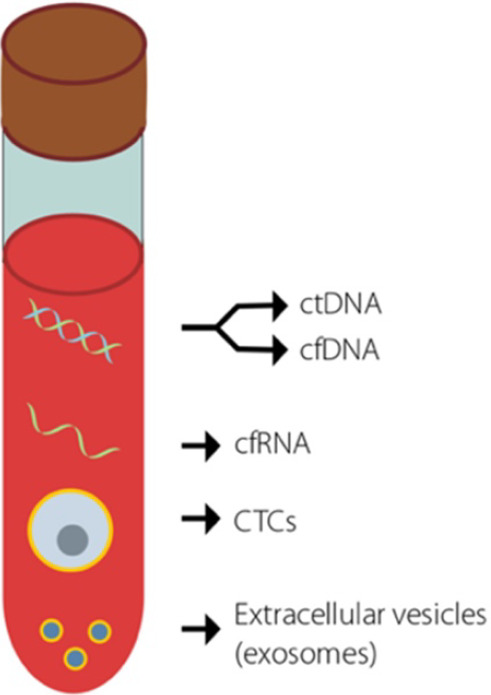
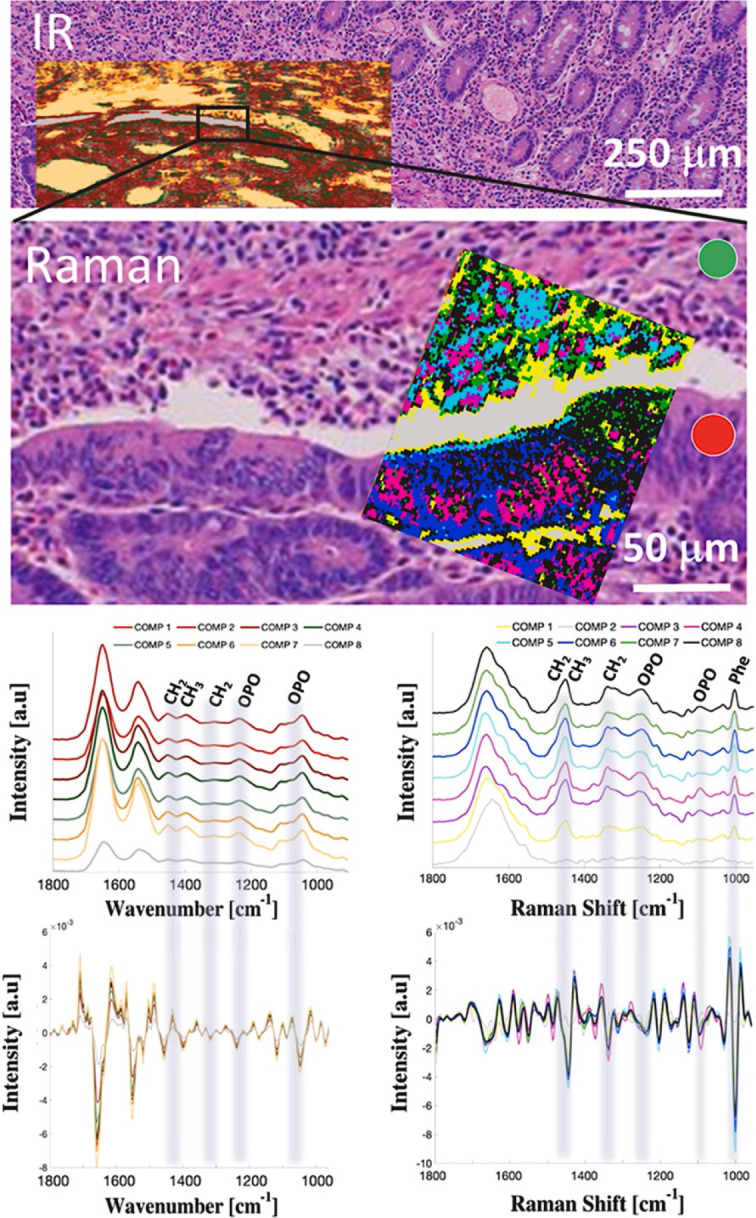
Keywords: Convolutional neural networks; DNA methylation; Liquid biopsy biomarkers; Pancreatic cancer diagnosis; Raman spectroscopy; Spectroscopic cancer diagnosis.
©The Author(s) 2023. Published by Baishideng Publishing Group Inc. All rights reserved.
Conflict of interest statement
Conflict-of-interest statement: All the authors report no relevant conflicts of interest for this article.
Figures


Similar articles
-
Variabilities in global DNA methylation and β-sheet richness establish spectroscopic landscapes among subtypes of pancreatic cancer.Eur J Nucl Med Mol Imaging. 2023 May;50(6):1792-1810. doi: 10.1007/s00259-023-06121-7. Epub 2023 Feb 9. Eur J Nucl Med Mol Imaging. 2023. PMID: 36757432 Free PMC article.
-
Current Pathology Model of Pancreatic Cancer.Cancers (Basel). 2022 May 7;14(9):2321. doi: 10.3390/cancers14092321. Cancers (Basel). 2022. PMID: 35565450 Free PMC article. Review.
-
Untargeted LC-HRMS-Based Metabolomics for Searching New Biomarkers of Pancreatic Ductal Adenocarcinoma: A Pilot Study.SLAS Discov. 2017 Apr;22(4):348-359. doi: 10.1177/1087057116671490. Epub 2016 Sep 27. SLAS Discov. 2017. PMID: 27655283
-
Liquid biopsy in pancreatic ductal adenocarcinoma: current status of circulating tumor cells and circulating tumor DNA.Mol Oncol. 2019 Aug;13(8):1623-1650. doi: 10.1002/1878-0261.12537. Epub 2019 Jul 30. Mol Oncol. 2019. PMID: 31243883 Free PMC article. Review.
-
SST gene hypermethylation acts as a pan-cancer marker for pancreatic ductal adenocarcinoma and multiple other tumors: toward its use for blood-based diagnosis.Mol Oncol. 2020 Jun;14(6):1252-1267. doi: 10.1002/1878-0261.12684. Epub 2020 Apr 14. Mol Oncol. 2020. PMID: 32243066 Free PMC article.
Cited by
-
Combined analytical approach empowers precise spectroscopic interpretation of subcellular components of pancreatic cancer cells.Anal Bioanal Chem. 2023 Dec;415(29-30):7281-7295. doi: 10.1007/s00216-023-04997-w. Epub 2023 Oct 31. Anal Bioanal Chem. 2023. PMID: 37906289 Free PMC article.
-
Variabilities in global DNA methylation and β-sheet richness establish spectroscopic landscapes among subtypes of pancreatic cancer.Eur J Nucl Med Mol Imaging. 2023 May;50(6):1792-1810. doi: 10.1007/s00259-023-06121-7. Epub 2023 Feb 9. Eur J Nucl Med Mol Imaging. 2023. PMID: 36757432 Free PMC article.
References
-
- Sung H, Ferlay J, Siegel RL, Laversanne M, Soerjomataram I, Jemal A, Bray F. Global Cancer Statistics 2020: GLOBOCAN Estimates of Incidence and Mortality Worldwide for 36 Cancers in 185 Countries. CA Cancer J Clin. 2021;71:209–249. - PubMed
-
- Takaori K, Bassi C, Biankin A, Brunner TB, Cataldo I, Campbell F, Cunningham D, Falconi M, Frampton AE, Furuse J, Giovannini M, Jackson R, Nakamura A, Nealon W, Neoptolemos JP, Real FX, Scarpa A, Sclafani F, Windsor JA, Yamaguchi K, Wolfgang C, Johnson CD IAP/EPC study group on the clinical managements of pancreatic cancer. International Association of Pancreatology (IAP)/European Pancreatic Club (EPC) consensus review of guidelines for the treatment of pancreatic cancer. Pancreatology. 2016;16:14–27. - PubMed
Publication types
MeSH terms
Substances
LinkOut - more resources
Full Text Sources
Medical
