

Epsilon-caprolactone-modified polyethylenimine as a genetic vehicle for stem cell-based bispecific antibody and exosome synergistic therapy
- PMID: 36683744
- PMCID: PMC9847525
- DOI: 10.1093/rb/rbac090
Epsilon-caprolactone-modified polyethylenimine as a genetic vehicle for stem cell-based bispecific antibody and exosome synergistic therapy
Abstract
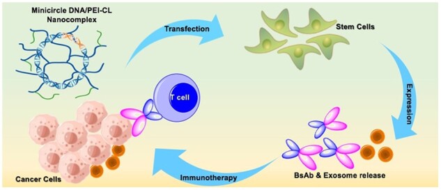
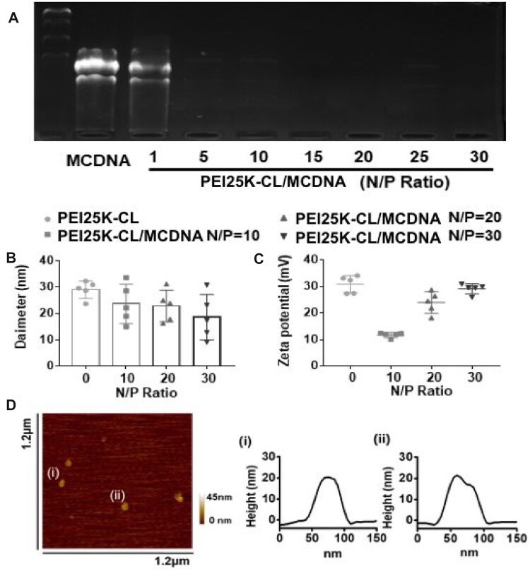
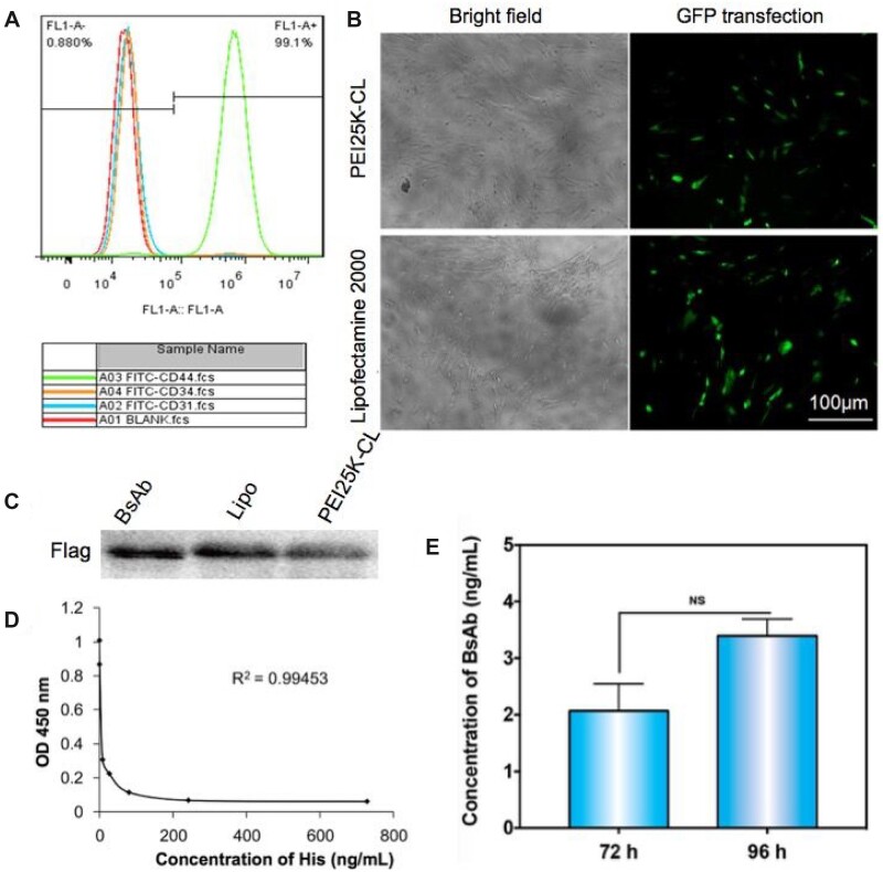
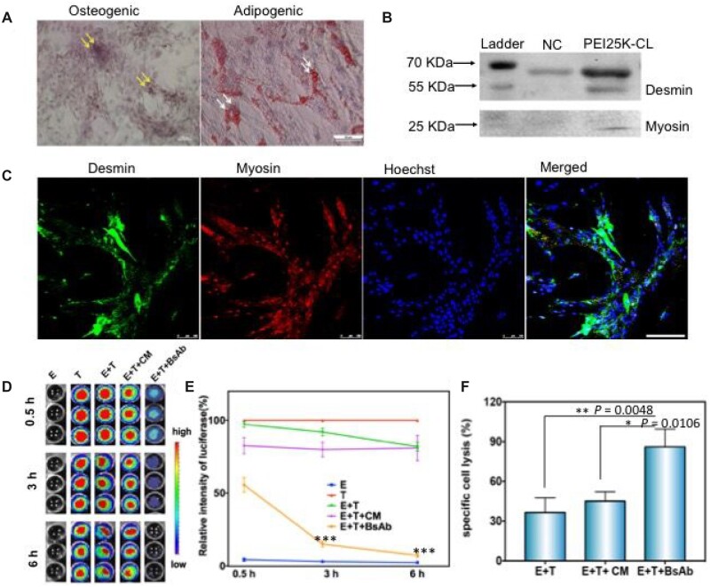
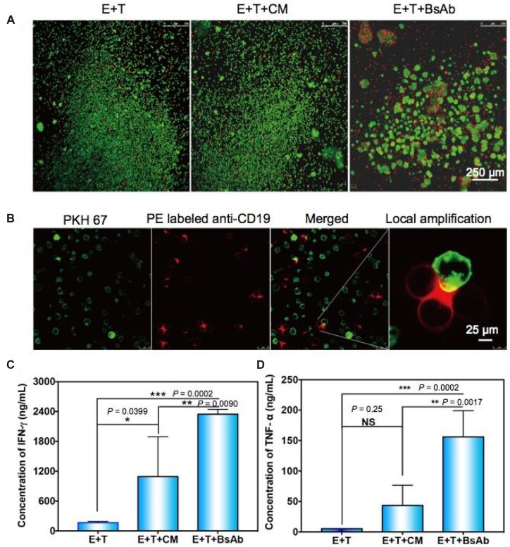
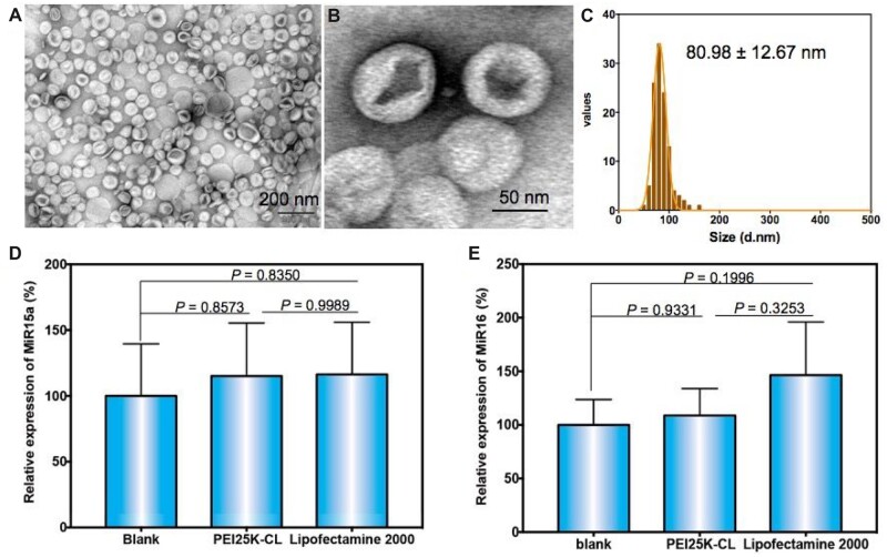
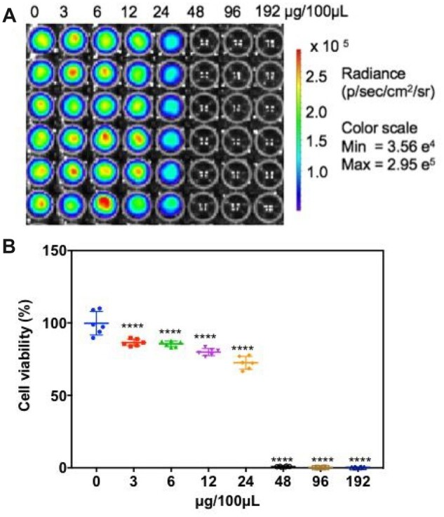
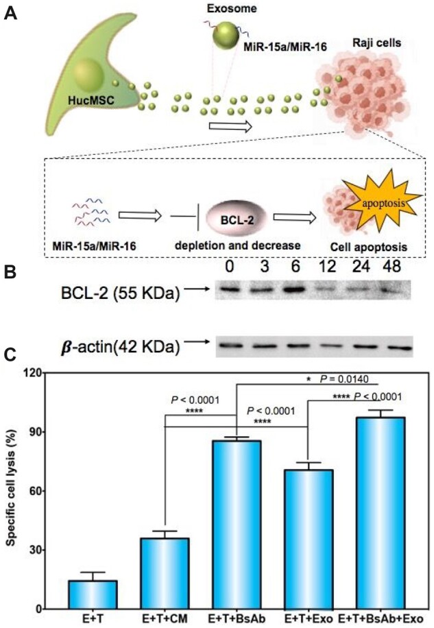
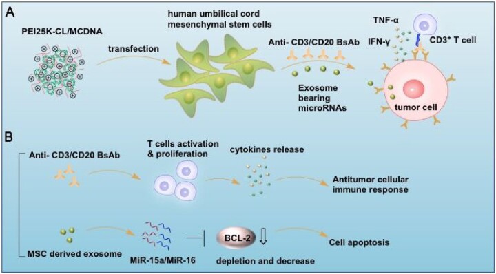
Bispecific antibodies (BsAb) have gained significant momentum in clinical application. However, the rapid enzymolysis and metabolism of protein drugs usually induce short circulation in vivo, and developing an efficient protein delivery system still is a bottleneck. Mesenchymal stem cells (MSCs) have become an attractive therapeutic carrier for cancers. Genetic modification enables MSCs to express and secrete specific proteins, which is essential for therapeutic efficacy. However, efficient gene transfer into MSCs is still a challenge. In this study, we applied epsilon-caprolactone-modified polyethylenimine (PEI-CL) as an efficacy carrier for plasmid transfection into MSC that served as in situ 'cell factory' for anti-CD3/CD20 BsAb preparation. Herein, the PEI-CL encapsulates the minicircle plasmid and mediates cell transfection efficiently. Thus, the anti-CD3/CD20 BsAb is secreted from MSC and recruited T cell, resulting in highly sensitive cytotoxicity in the human B-cell lymphoma. Furthermore, these stem cells produce exosomes bearing MiR-15a/MiR-16, which could negatively regulate cancer's oncogenes BCL-2 for adjuvant therapy. Meanwhile, high immunologic factors like tumor necrosis factor-α and interferon-γ are generated and enhance immunotherapy efficacy. The engineered MSCs are demonstrated as an efficient route for BsAb production, and these bioactive components contribute to synergistic therapy, which would be an innovative treatment.
Keywords: bispecific antibody; exosome; polyethylenimine; stem cell.
© The Author(s) 2022. Published by Oxford University Press.
Figures
Similar articles
-
Mesenchymal stromal cells as vehicles of tetravalent bispecific Tandab (CD3/CD19) for the treatment of B cell lymphoma combined with IDO pathway inhibitor D-1-methyl-tryptophan.J Hematol Oncol. 2017 Feb 23;10(1):56. doi: 10.1186/s13045-017-0397-z. J Hematol Oncol. 2017. PMID: 28228105 Free PMC article.
-
Polylysine-modified polyethylenimine polymer can generate genetically engineered mesenchymal stem cells for combinational suicidal gene therapy in glioblastoma.Acta Biomater. 2018 Oct 15;80:144-153. doi: 10.1016/j.actbio.2018.09.015. Epub 2018 Sep 15. Acta Biomater. 2018. PMID: 30223091
-
BL-01, an Fc-bearing, tetravalent CD20 × CD5 bispecific antibody, redirects multiple immune cells to kill tumors in vitro and in vivo.Cytotherapy. 2022 Feb;24(2):161-171. doi: 10.1016/j.jcyt.2021.07.012. Epub 2021 Sep 17. Cytotherapy. 2022. PMID: 34538717
-
Treatment of Human B-Cell Lymphomas Using Minicircle DNA Vector Expressing Anti-CD3/CD20 in a Mouse Model.Hum Gene Ther. 2017 Feb;28(2):216-225. doi: 10.1089/hum.2016.122. Epub 2016 Nov 1. Hum Gene Ther. 2017. PMID: 27802782 Review.
-
Stem Cell Mimicking Nanoencapsulation for Targeting Arthritis.Int J Nanomedicine. 2021 Dec 31;16:8485-8507. doi: 10.2147/IJN.S334298. eCollection 2021. Int J Nanomedicine. 2021. PMID: 35002240 Free PMC article. Review.
Cited by
-
Mesenchymal Stem Cell-Derived Extracellular Vesicles in Cancer Therapy Resistance: from Biology to Clinical Opportunity.Int J Biol Sci. 2024 Jan 1;20(1):347-366. doi: 10.7150/ijbs.88500. eCollection 2024. Int J Biol Sci. 2024. PMID: 38164177 Free PMC article. Review.
-
Extracellular Vesicles from Mesenchymal Stem Cells: Potential as Therapeutics in Metabolic Dysfunction-Associated Steatotic Liver Disease (MASLD).Biomedicines. 2024 Dec 14;12(12):2848. doi: 10.3390/biomedicines12122848. Biomedicines. 2024. PMID: 39767754 Free PMC article. Review.
-
Exosomes-based dual drug-loaded nanocarrier for targeted and multiple proliferative vitreoretinopathy therapy.Regen Biomater. 2024 Jun 29;11:rbae081. doi: 10.1093/rb/rbae081. eCollection 2024. Regen Biomater. 2024. PMID: 39040514 Free PMC article.
-
Advancements in engineered mesenchymal stem cell exosomes for chronic lung disease treatment.J Transl Med. 2023 Dec 9;21(1):895. doi: 10.1186/s12967-023-04729-9. J Transl Med. 2023. PMID: 38071321 Free PMC article. Review.
References
-
- Douglass J, Hsiue EH, Mog BJ, Hwang MS, DiNapoli SR, Pearlman AH, Miller MS, Wright KM, Azurmendi PA, Wang Q, Paul S, Schaefer A, Skora AD, Molin MD, Konig MF, Liu Q, Watson E, Murphy LY, Pardoll MB, Bettegowda DM, Papadopoulos C, Gabelli N, Kinzler SB, Vogelstein KW, Zhou B.. Bispecific antibodies targeting mutant RAS neoantigens. Sci Immunol 2021;6:eabd5515. - PMC - PubMed
-
- Xu G, Luo Y, Wang H, Wang Y, Liu B, Wei J.. Therapeutic bispecific antibodies against intracellular tumor antigens. Cancer Lett 2022;538:215699. - PubMed
-
- Labrijn AF, Meesters JI, de Goeij BE, van den Bremer ET, Neijssen J, van Kampen MD, Strumane K, Verploegen S, Kundu A, Gramer MJ, van Berkel PH, van de Winkel JG, Schuurman J, Parren PW.. Efficient generation of stable bispecific IgG1 by controlled Fab-arm exchange. Proc Natl Acad Sci USA 2013;110:5145–50. - PMC - PubMed
LinkOut - more resources
Full Text Sources
