

Micropatterned composite membrane guides oriented cell growth and vascularization for accelerating wound healing
- PMID: 36683746
- PMCID: PMC9847515
- DOI: 10.1093/rb/rbac108
Micropatterned composite membrane guides oriented cell growth and vascularization for accelerating wound healing
Abstract
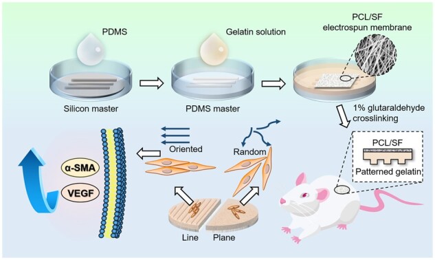
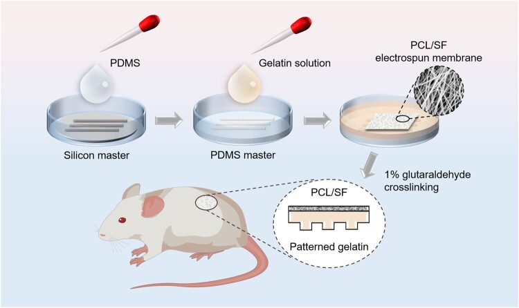
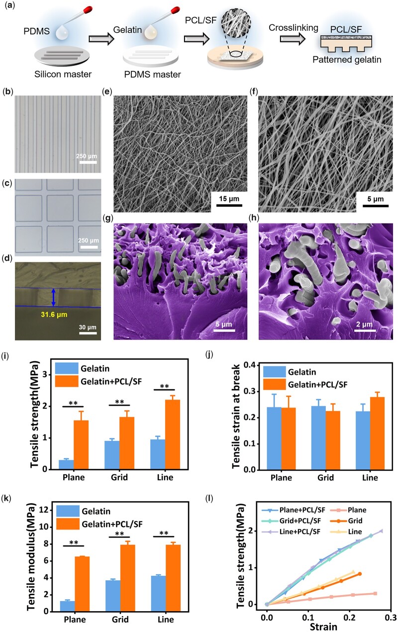
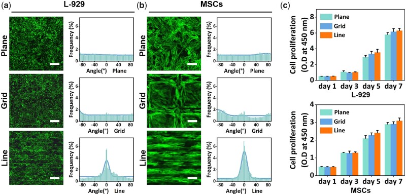
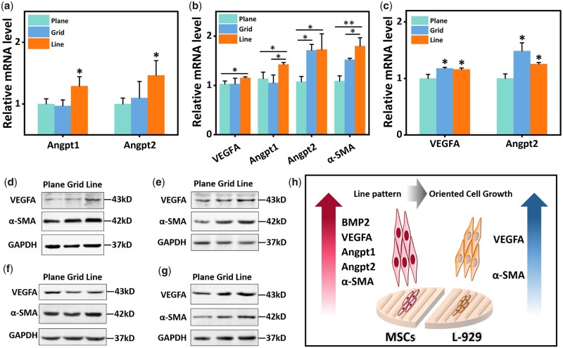
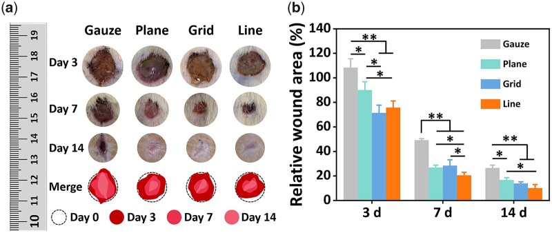
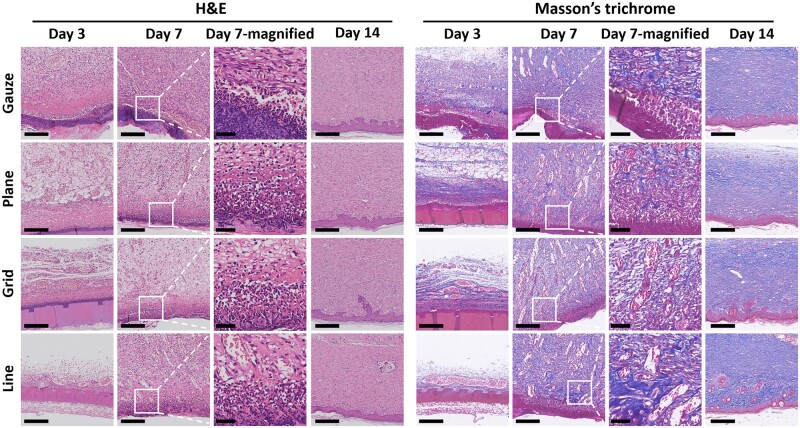
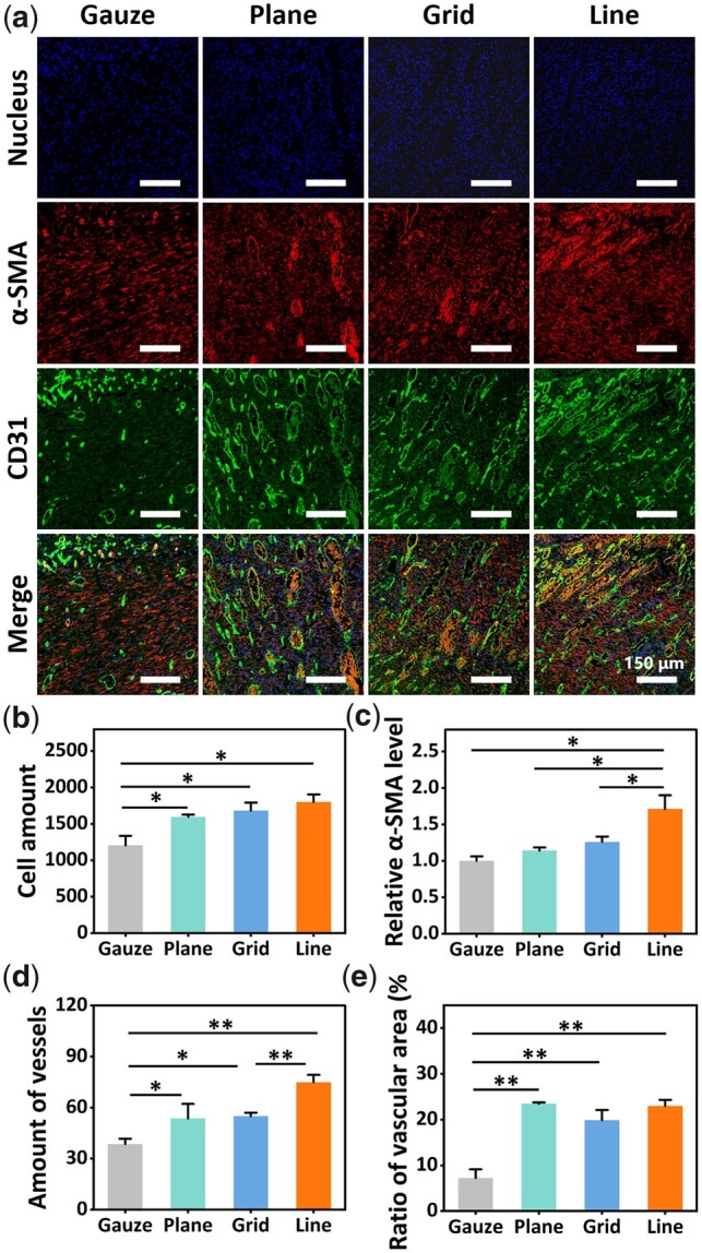
Skin defect is common in daily life, but repairing large skin defects remains a challenge. Using biomaterials to deliver biochemical or physical factors to promote skin tissue regeneration is of great significance for accelerating wound healing. Specific surface micropatterns on biomaterials could affect cell behavior and tissue regeneration. However, few studies have focused on the construction of wound healing biomaterials with surface micropatterns and their role in skin tissue regeneration. In the present study, gelatin-polycaprolactone/silk fibroin composite membranes with different micropatterns were fabricated by photolithography, including line, grid and plane micropatterns. In vitro cell experiments demonstrated that the line micropattern on the composite membrane could guide cell-oriented growth, and more importantly, promote the expression of angiogenesis-related markers and α-smooth muscle actin (α-SMA) at both gene level and protein level. In the rat full-thickness skin defect model, the composite membrane with line micropatterns increased α-SMA production and neovascularization in wounds, leading to accelerated wound contraction and healing. The current study not only suggests that composite membranes with specific micropatterns can be promising wound repair materials but also provides new insights into the importance of biomaterial surface topology for tissue regeneration.
Keywords: cell behavior; gelatin; polymer biomaterials; topology; wound healing.
© The Author(s) 2022. Published by Oxford University Press.
Figures
Similar articles
-
Human urine-derived stem cells in combination with polycaprolactone/gelatin nanofibrous membranes enhance wound healing by promoting angiogenesis.J Transl Med. 2014 Oct 2;12:274. doi: 10.1186/s12967-014-0274-2. J Transl Med. 2014. PMID: 25274078 Free PMC article.
-
Photo-crosslinkable amniotic membrane hydrogel for skin defect healing.Acta Biomater. 2021 Apr 15;125:197-207. doi: 10.1016/j.actbio.2021.02.043. Epub 2021 Mar 4. Acta Biomater. 2021. PMID: 33676048
-
A conducive bioceramic/polymer composite biomaterial for diabetic wound healing.Acta Biomater. 2017 Sep 15;60:128-143. doi: 10.1016/j.actbio.2017.07.020. Epub 2017 Jul 14. Acta Biomater. 2017. PMID: 28713016
-
Silk biomaterials in wound healing and skin regeneration therapeutics: From bench to bedside.Acta Biomater. 2020 Feb;103:24-51. doi: 10.1016/j.actbio.2019.11.050. Epub 2019 Dec 2. Acta Biomater. 2020. PMID: 31805409 Review.
-
Silk Fibroin Biomaterials and Their Beneficial Role in Skin Wound Healing.Biomolecules. 2022 Dec 12;12(12):1852. doi: 10.3390/biom12121852. Biomolecules. 2022. PMID: 36551280 Free PMC article. Review.
Cited by
-
3D Printing of Tough Hydrogel Scaffolds with Functional Surface Structures for Tissue Regeneration.Nanomicro Lett. 2024 Sep 29;17(1):27. doi: 10.1007/s40820-024-01524-z. Nanomicro Lett. 2024. PMID: 39342523 Free PMC article.
-
Spatial confinement modulates endothelial cell behavior and traction force in 3D hydrogel microgrooves.Mater Today Bio. 2024 Apr 30;26:101074. doi: 10.1016/j.mtbio.2024.101074. eCollection 2024 Jun. Mater Today Bio. 2024. PMID: 38736613 Free PMC article.
-
Biomaterials for neuroengineering: applications and challenges.Regen Biomater. 2025 Feb 21;12:rbae137. doi: 10.1093/rb/rbae137. eCollection 2025. Regen Biomater. 2025. PMID: 40007617 Free PMC article. Review.
-
Advances in tissue engineering and biofabrication for in vitro skin modeling.Bioprinting. 2023 Nov;35:e00306. doi: 10.1016/j.bprint.2023.e00306. Epub 2023 Sep 1. Bioprinting. 2023. PMID: 38645432 Free PMC article.
-
Cancer cell response to extrinsic and intrinsic mechanical cue: opportunities for tumor apoptosis strategies.Regen Biomater. 2024 Feb 20;11:rbae016. doi: 10.1093/rb/rbae016. eCollection 2024. Regen Biomater. 2024. PMID: 38476678 Free PMC article. Review.
References
-
- Winter GD. Formation of the scab and the rate of epithelization of superficial wounds in the skin of the young domestic pig. J Wound Care 1995;193:366–7. - PubMed
LinkOut - more resources
Full Text Sources
