

Biological properties of self-assembled nanofibers of elastin-like block polypeptides for tissue-engineered vascular grafts: platelet inhibition, endothelial cell activation and smooth muscle cell maintenance
- PMID: 36683748
- PMCID: PMC9845521
- DOI: 10.1093/rb/rbac111
Biological properties of self-assembled nanofibers of elastin-like block polypeptides for tissue-engineered vascular grafts: platelet inhibition, endothelial cell activation and smooth muscle cell maintenance
Abstract
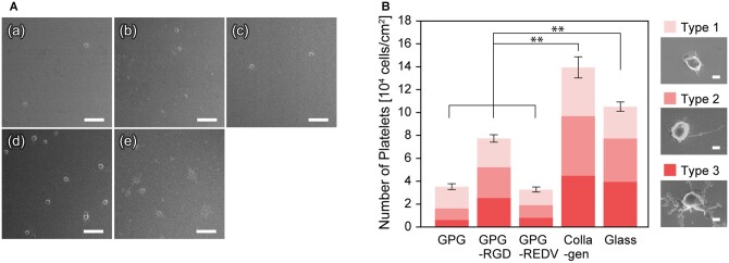
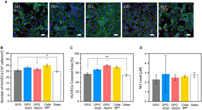
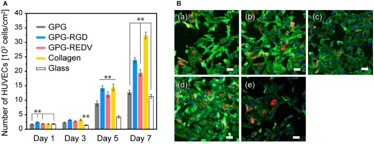
Strategic materials design is essential for the development of small-diameter, tissue-engineered vascular grafts. Self-assembled nanofibers of elastin-like polypeptides represent promising vascular graft components as they replicate the organized elastin structure of native blood vessels. Further, the bioactivity of nanofibers can be modified by the addition of functional peptide motifs. In the present study, we describe the development of a novel nanofiber-forming elastin-like polypeptide (ELP) with an arginine-glutamic acid-aspartic acid-valine (REDV) sequence. The biological characteristics of the REDV-modified ELP nanofibers relevant to applications in vascular grafting were compared to ELP without ligands for integrin, ELP with arginine-glycine-aspartic acid (RGD) sequence, collagen and cell culture glass. Among them, REDV-modified ELP nanofibers met the preferred biological properties for vascular graft materials, i.e. (i) inhibition of platelet adhesion and activation, (ii) endothelial cell adhesion and proliferation and (iii) maintenance of smooth muscle cells in a contractile phenotype to prevent cell overgrowth. The results indicate that REDV-modified ELP nanofibers represent promising candidates for the further development of small-diameter vascular grafts.
Keywords: elastin-like polypeptide; endothelial cell; nanofiber; platelet; smooth muscle cell.
© The Author(s) 2022. Published by Oxford University Press.
Figures


References
-
- Hiob MA, She S, Muiznieks LD, Weiss AS.. Biomaterials and modifications in the development of small-diameter vascular grafts. ACS Biomater Sci Eng 2017;3:712–23. - PubMed
-
- Gupta P, Mandal BB.. Tissue-engineered vascular grafts: emerging trends and technologies. Adv Funct Materials 2021;31:2100027.
LinkOut - more resources
Full Text Sources
Miscellaneous
