. 2022 Oct 26:10:rbac085.
doi: 10.1093/rb/rbac085. eCollection 2023.

Integrated printed BDNF-stimulated HUCMSCs-derived exosomes/collagen/chitosan biological scaffolds with 3D printing technology promoted the remodelling of neural networks after traumatic brain injury

Affiliations

Integrated printed BDNF-stimulated HUCMSCs-derived exosomes/collagen/chitosan biological scaffolds with 3D printing technology promoted the remodelling of neural networks after traumatic brain injury

Xiaoyin Liu et al. Regen Biomater. .

Abstract

The restoration of nerve dysfunction after traumatic brain injury (TBI) faces huge challenges due to the limited self-regenerative abilities of nerve tissues. In situ inductive recovery can be achieved utilizing biological scaffolds combined with endogenous human umbilical cord mesenchymal stem cells (HUCMSCs)-derived exosomes (MExos). In this study, brain-derived neurotrophic factor-stimulated HUCMSCs-derived exosomes (BMExos) were composited with collagen/chitosan by 3D printing technology. 3D-printed collagen/chitosan/BMExos (3D-CC-BMExos) scaffolds have excellent mechanical properties and biocompatibility. Subsequently, in vivo experiments showed that 3D-CC-BMExos therapy could improve the recovery of neuromotor function and cognitive function in a TBI model in rats. Consistent with the behavioural recovery, the results of histomorphological tests showed that 3D-CC-BMExos therapy could facilitate the remodelling of neural networks, such as improving the regeneration of nerve fibres, synaptic connections and myelin sheaths, in lesions after TBI.

Keywords: BDNF; chitosan; collagen; exosomes; mesenchymal stem cell; traumatic brain injury.


Figures

None
Graphical abstract
Figure 1.
Figure 1.
Characterization of Exos derived from HUCMSCs under BDNF conditions and non-BDNF conditions. (A) There were no significant alterations in the morphology and growth of HUCMSCs when BDNF was used to stimulate the culture medium of MSCs. (B) Morphological characteristics of MExos and BMExos under TEM. (C) Exosomal-specific protein markers CD9 and CD63 investigated by western blotting. (D) The diameter of MExos and BMExos measured using NanosizerTM technology. (E) The concentration of MExos was 0.97 ± 0.31 µg/ml and the concentration of BMExos was 1.13 ± 0.36 µg/ml, no significant differences were found. TEM, transmission electron microscopy.
Figure 2.
Figure 2.
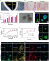
Characteristics of composite scaffolds. (AD) Representative images of 3D-CC-BMExos under general observation (A), H&E staining (B) and SEM (C and D). The pores of 3D-CC-BMExos were well connected to each other and the BMExos were evenly distributed in the 3D-CC-BMExos scaffold (D). (E) Representative 3D immunofluorescence images showed the distribution of BMExos in 3D-CC-BMExos. (F) Degradation rate of 3D-CC-BMExos prepared with different collagen/chitosan mass ratios at 2, 4, 6 and 8 weeks after transplantation. (G and H) Compared with CC scaffolds, 3D-printed composite scaffolds showed lower water absorption ratio and higher porosity ratio. (I) Cumulative release profile of BMExos from the CC-BMExos and 3D-CC-BMExos within 14 days. (J and K) Representative images of F-actin/PKH26-labelled BMExos immunofluorescence staining in HUCMSCs (J) and NSCs (K). All data were expressed as mean ± SD; *P <0.05, **P <0.01 vs CC; #P <0.05, ##P <0.01 vs CC-BMExos; SEM, scanning electron microscope.
Figure 3.
Figure 3.
Biocompatibility test of scaffolds. (A and B) Representative images of HUCMSCs cocultured with 3D-CC-MExos or 3D-CC-BMExos under phase contrast microscopy (A) and H&E staining (B). (C) MTT assay of HUCMSCs cocultured with 3D-CC-MExos and 3D-CC-BMExos. (D) NSCs were spherical under an optical microscope. (E) Representative immunofluorescence images of NSCs stained with a nestin antibody (green). (F) Cell adhesion rates of NSCs at different time points after coculture of NSCs with scaffolds. (G) MTT assay of NSCs cocultured with 3D-CC-MExos and 3D-CC-BMExos. (HL) Representative image of F-actin (H), NF (I), NeuN (J), GAP42 (K) and GFAP (L) immunofluorescence staining at 7 days after coculture. All data were expressed as mean ± SD; *P <0.05, **P <0.01 vs 3D-CC-MExos.
Figure 4.
Figure 4.
The recovery of cognitive function and sensorimotor function among the four groups. (A) Representative images of the search route in the spatial learning stage. The yellow arrow represents the platform, and the zone labelled by the red five-pointed star is the target zone. (B) The analysis of escape latency in the spatial learning stage. (C and D) Analysis of the number of site crossings (C) and the time ratio in the target zone (D) in the spatial memory stage. (E) mNSS assessment at different time points after TBI. All data were collected as mean ± SD; *P <0.05, **P <0.01 vs TBI group; #P <0.05, ##P <0.01 vs 3D-CC-MExos group.
Figure 5.
Figure 5.
3D-CC-BMExos therapy promoted histomorphological recovery at the injury site after TBI. (A) Representative H&E-stained images of brain slices among the four groups showing the differences in histomorphology at 2 months after TBI. (B) Representative Bielschowsky’s silver staining images of brain slices among the four groups showing the differences in nerve fibre alterations at 2 months after TBI. (C) Representative Nissl staining images of brain slices among the four groups showing the differences in the numbers of neuronal cell bodies at 2 months after TBI. (D) General observation of cavity after TBI among the four groups. (E) Quantitative measurement of cavity area per field at the defect site. (F) Quantitative measurement of bielschowsky’s silver staining area per field at the defect site. (G) Quantitative measurement of nissl staining area per field at the defect site. All data were expressed as mean ± SD; *P <0.05, **P <0.01 vs TBI group; ##P <0.01 vs 3D-CC-MExos group.
Figure 6.
Figure 6.
The recovery of nerve fibres, myelin sheaths and neurons at the injury site of TBI. (A) Representative immunofluorescence images of nerve fibres, myelin sheaths and neurons in the injury site. (B) Representative TEM images of myelin sheaths and neurons. (C–E) The quantification of NF+ cells (C), MBP+ cells (D) and NeuN+ cells (E) in the injury site among the four groups. All data were expressed as mean ± SD; **P <0.01 vs TBI group; #P <0.05, ##P <0.01 vs 3D-CC-MExos group; TEM, transmission electron microscope.
Figure 7.
Figure 7.
The recovery of synapses and axons at the injury site of TBI. (A) Representative immunofluorescence images of axons and synapses in the injury site. (B and C) Quantification of the MAP2+ cells (B) and SYP+ cells (C) in the injury site among the four groups. All data were expressed as mean ± SD; **P <0.01 vs TBI group; ##P <0.01 vs 3D-CC-MExos group.
Figure 8.
Figure 8.
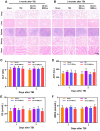
In vivo toxicity of transplanted composite scaffolds. (A and B) H&E staining of the lung, liver, spleen, kidney and heart at 1 month and 2 months after TBI among the four groups. (CF) There were no significant differences in lab blood tests of ALT, AST, CR and UREA among the four groups. ALT, alanine aminotransferase; AST, aspartate aminotransferase; CR, serum creatinine; UREA, blood urea nitrogen.

Similar articles

Cited by

References

    1. Fu S, Zhao S, Chen H, Yang W, Xia X, Xu X, Liang Z, Feng X, Wang Z, Ai P, Ding L, Cai Q, Wang Y, Zhang Y, Zhu J, Zhang B, Zheng JC.. Insulin-incubated palladium clusters promote recovery after brain injury. J Nanobiotechnology 2022;20:299. - PMC - PubMed
    1. Tian L, Prabhakaran MP, Ramakrishna S.. Strategies for regeneration of components of nervous system: scaffolds, cells and biomolecules. Regen Biomater 2015;2:31–45. - PMC - PubMed
    1. Qian F, Han Y, Han Z, Zhang D, Zhang L, Zhao G, Li S, Jin G, Yu R, Liu H.. In situ implantable, post-trauma microenvironment-responsive, ROS depletion hydrogels for the treatment of traumatic brain injury. Biomaterials 2021;270:120675. - PubMed
    1. Ma S, Zhou J, Huang T, Zhang Z, Xing Q, Zhou X, Zhang K, Yao M, Cheng T, Wang X, Wen X, Guan F.. Sodium alginate/collagen/stromal cell-derived factor-1 neural scaffold loaded with BMSCs promotes neurological function recovery after traumatic brain injury. Acta Biomater 2021;131:185–97. - PubMed
    1. Li J, Zhang D, Guo S, Zhao C, Wang L, Ma S, Guan F, Yao M.. Dual-enzymatically cross-linked gelatin hydrogel promotes neural differentiation and neurotrophin secretion of bone marrow-derived mesenchymal stem cells for treatment of moderate traumatic brain injury. Int J Biol Macromol 2021;187:200–13. - PubMed