. 2022 Dec 13:10:rbac102.
doi: 10.1093/rb/rbac102. eCollection 2023.

NIR-II live imaging study on the degradation pattern of collagen in the mouse model

Affiliations

NIR-II live imaging study on the degradation pattern of collagen in the mouse model

Huizhu Li et al. Regen Biomater. .

Abstract

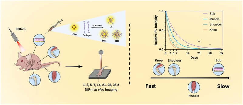
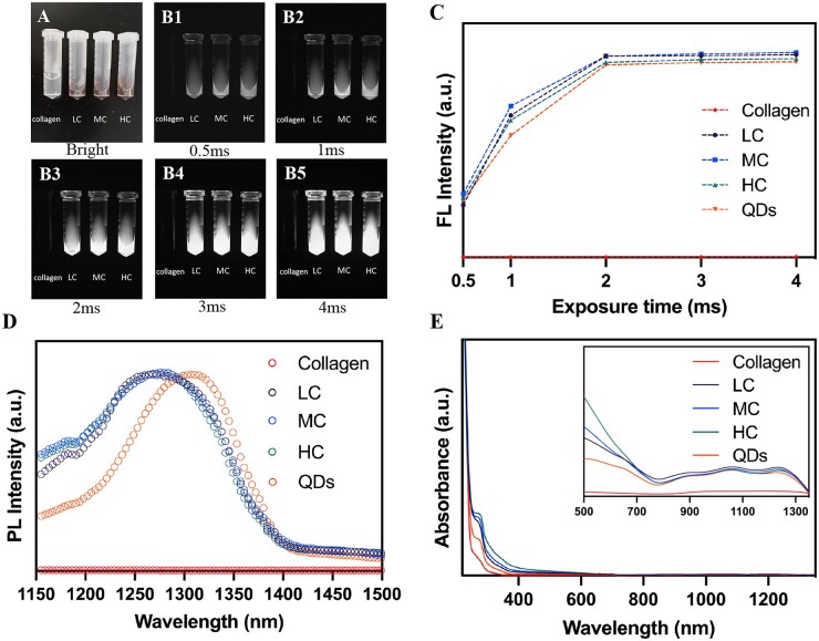
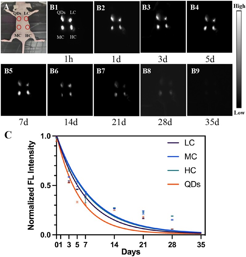
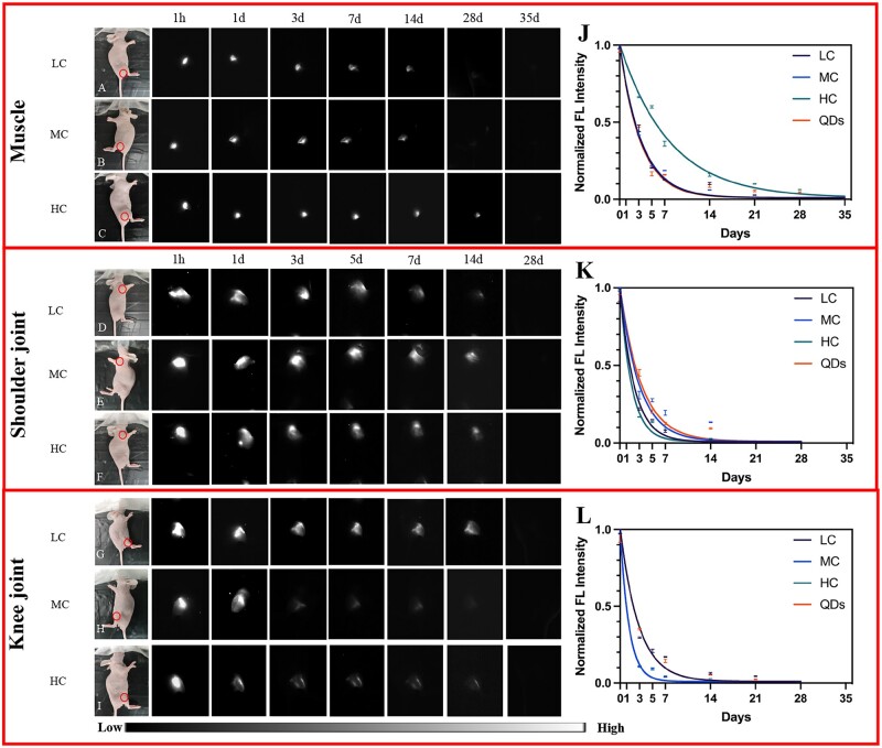
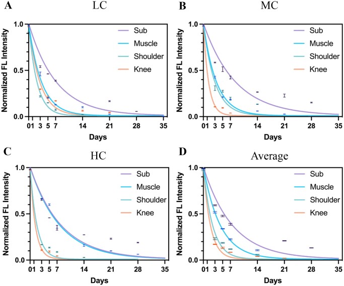
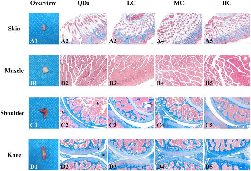
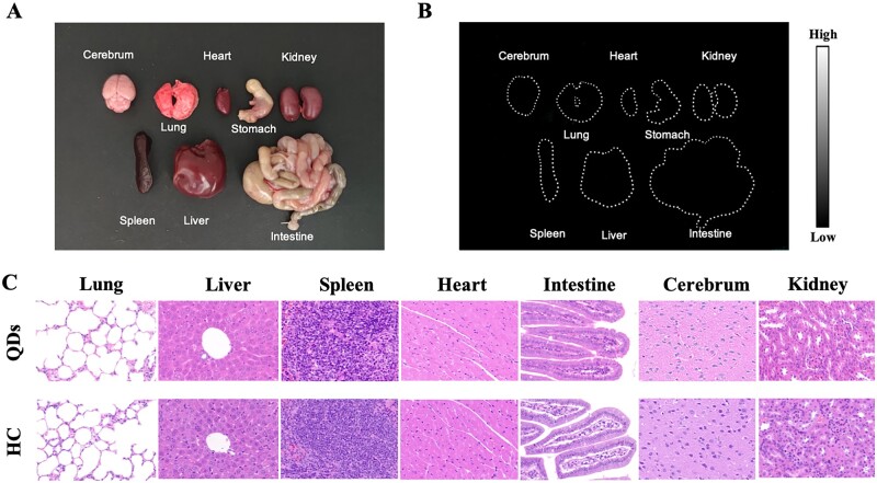
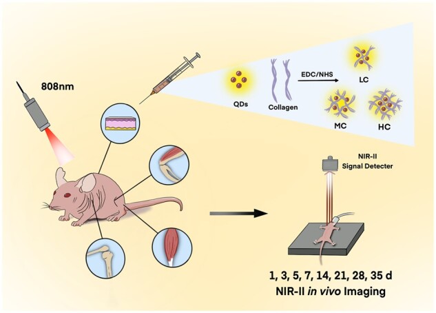
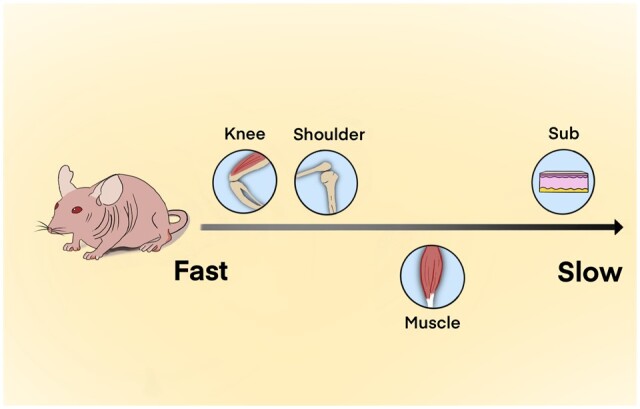
The degradation of collagen in different body parts is a critical point for designing collagen-based biomedical products. Here, three kinds of collagens labeled by second near-infrared (NIR-II) quantum dots (QDs), including collagen with low crosslinking degree (LC), middle crosslinking degree (MC) and high crosslinking degree (HC), were injected into the subcutaneous tissue, muscle and joints of the mouse model, respectively, in order to investigate the in vivo degradation pattern of collagen by NIR-II live imaging. The results of NIR-II imaging indicated that all tested collagens could be fully degraded after 35 days in the subcutaneous tissue, muscle and joints of the mouse model. However, the average degradation rate of subcutaneous tissue (k = 0.13) and muscle (k = 0.23) was slower than that of the joints (shoulder: k = 0.42, knee: k = 0.55). Specifically, the degradation rate of HC (k = 0.13) was slower than LC (k = 0.30) in muscle, while HC showed the fastest degradation rate in the shoulder and knee joints. In summary, NIR-II imaging could precisely identify the in vivo degradation rate of collagen. Moreover, the degradation rate of collagen was more closely related to the implanted body parts rather than the crosslinking degree of collagen, which was slower in the subcutaneous tissue and muscle compared to the joints in the mouse model.

Keywords: NIR-II live imaging; collagen; crosslinking degree; degradation rate; in vivo.


Figures

None
Graphical abstract
Figure 1.
Figure 1.
Characterization of QDs-labeled collagen. (A) Bright-field and (B1-B5) NIR-II images of collagen, LC, MC and HC at different exposure times. Corresponding FL intensity (C), PL spectrum (D) and absorbance spectrum (E) of collagen, LC, MC, HC and QDs under 808-nm excitation.
Figure 2.
Figure 2.
Collagen degradation in subcutaneous tissue. (A) Bright-field image of the subcutaneously injected mouse. (B1-B9) NIR-II fluorescence images were taken at 1 h, 1, 3, 5, 7, 14, 21, 28 and 35 days after injection of QDs, LC, MC and HC. (C) Corresponding FL intensity analysis (B) and colored solid curve representing the degradation rate of QDs, LC, MC and HC, respectively.
Figure 3.
Figure 3.
Collagen degradation in muscle, shoulder joint and knee joint. (A–C) Bright-field and NIR-II fluorescence images of LC (A), MC (B) and HC (C) degradation in muscle. (D–F) Bright-field and NIR-II fluorescence images of LC (D), MC (E) and HC (F) degradation in the shoulder joint. (G–I) Bright-field and NIR-II fluorescence images of LC (G), MC (H) and HC (I) degradation in the knee joint (circles: injection sites). (J–L) Corresponding normalized FL intensity analysis of collagen and degradation rate in muscle (J), shoulder joint (K) and knee joint (L).
Figure 4.
Figure 4.
LC, MC and HC degradation in different tissue. Normalized FL intensity and degradation rate of LC (A), MC (B) and HC (C) in subcutaneous tissue, muscle, shoulder joint and knee joint ranging from 1 h to 35 days. (D) Average normalized FL intensity and degradation rate of collagen in subcutaneous tissue, muscle, shoulder joint and knee joint, respectively.
Figure 5.
Figure 5.
Macroscopic and microscopic images of collected tissues after the signals disappeared. (A1–D1) Macroscopic images of collected skin (35 days), muscle (28 days for LC, MC; 35 days for HC), shoulder joint (28 days) and knee joint (28 days). (A2–D2) Micrographs of Masson’s trichrome-staining slices of tissues injected with (A2–D2) QDs, (A3–D3) LC, (A4–D4) MC and (A5–D5) HC. H, humerus; G, glenoid; F, femur; T, tibia; M, meniscus. Magnification: ×100 for (A2–A5), (B2–B5), (C2–C5), (D2–D5).
Figure 6.
Figure 6.
Biosafety of QDs-labeled collagen. (A) Bright-field image of major organs including cerebrum, lung, heart, stomach, kidney, spleen, liver and intestine from mouse injected with QDs-labeled collagen. (B) NIR-II fluorescence image of major organs. (C) HE staining of organs from QDs-injected mice and HC-injected mice. Magnification: ×400.
Scheme 1.
Scheme 1.
Schematic illustration of monitoring the collagen degradation in vivo.
Scheme 2.
Scheme 2.
Comparison of collagen degradation rate in different body parts of the mouse model.

Similar articles

Cited by

References

    1. Shekhter AB, Fayzullin AL, Vukolova MN, Rudenko TG, Osipycheva VD, Litvitsky PF.. Medical applications of collagen and collagen-based materials. Curr Med Chem 2019;26:506–16. - PubMed
    1. Grabska-Zielińska S, Sionkowska A, Carvalho Â, Monteiro FJ.. Biomaterials with potential use in bone tissue regeneration-collagen/chitosan/silk fibroin scaffolds cross-linked by EDC/NHS. Materials (Basel) 2021;14:1105. - PMC - PubMed
    1. An B, Lin YS, Brodsky B.. Collagen interactions: drug design and delivery. Adv Drug Deliv Rev 2016;97:69–84. - PubMed
    1. Makris EA, Gomoll AH, Malizos KN, Hu JC, Athanasiou KA.. Repair and tissue engineering techniques for articular cartilage. Nat Rev Rheumatol 2015;11:21–34. - PMC - PubMed
    1. Irawan V, Sung TC, Higuchi A, Ikoma T.. Collagen scaffolds in cartilage tissue engineering and relevant approaches for future development. Tissue Eng Regen Med 2018;15:673–97. - PMC - PubMed