. 2022 Nov 2:10:rbac088.
doi: 10.1093/rb/rbac088. eCollection 2023.

Fabrication of initial trabecular bone-inspired three-dimensional structure with cell membrane nano fragments

Affiliations

Fabrication of initial trabecular bone-inspired three-dimensional structure with cell membrane nano fragments

Koichi Kadoya et al. Regen Biomater. .

Abstract

The extracellular matrix of trabecular bone has a large surface exposed to the bone marrow and plays important roles such as hematopoietic stem cell niche formation and maintenance. In vitro reproduction of trabecular bone microenvironment would be valuable not only for developing a functional scaffold for bone marrow tissue engineering but also for understanding its biological functions. Herein, we analyzed and reproduced the initial stages of trabecular bone formation in mouse femur epiphysis. We identified that the trabecular bone formation progressed through the following steps: (i) partial rupture of hypertrophic chondrocytes; (ii) calcospherite formation on cell membrane nano fragments (CNFs) derived from the ruptured cells; and (iii) calcospherite growth and fusion to form the initial three-dimensional (3D) structure of trabecular bones. For reproducing the initial trabecular bone formation in vitro, we collected CNFs from cultured cells and used as nucleation sites for biomimetic calcospherite formation. Strikingly, almost the same 3D structure of the initial trabecular bone could be obtained in vitro by using additional CNFs as a binder to fuse biomimetic calcospherites.

Keywords: bone tissue synthesis; calcospherites; cell membrane nano fragments; three dimensionalization; trabecular bone.


Figures

None
Graphical abstract
Figure 1.
Figure 1.
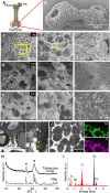
Analysis of the initial stages of trabecular bone formation in mouse femur epiphysis. (A) Schematic design of mouse femur. Circle indicates the epiphysis. (B) Secondary electron image of the initially formed trabecular bone at postnatal Day 8 (P8) after NaClO treatment for removal of all organic components. (C–E) Secondary electron images of the trabecular bones at (C) P8, (D) P10 and (E) P12 observed at different magnifications. (F) and (G) Backscattered electron images of a cross section near the front of the trabecular bone at P8. Right panel in (F) shows a higher magnified image of the area within the yellow squares in the left panel, and arrowheads indicate the calcospherites. Whitish multiform materials in the vicinity of the calcospherites would be rich in osmium-stained lipids of cell membrane fragments [14]. In the left panel in (G) taken at lower magnification, the fusion of calcospherites could be observed on the right upper area. Note that after calcospherite fusion, the trabecular bone became compact (left down area). Right panels in (G) indicate the localization of Ca and P in the trabecular bone. (H) XRD analysis showing the low crystalline HAp in the initially formed trabecular bone at P8. Commercially available HAp was used as a reference. (I) EDS analysis showing high peaks for Ca and P in the native trabecular bone at P8.
Figure 2.
Figure 2.
In vitro fabrication of calcospherites with cell nanofragments (CNFs). (A) Schematic design showing the protocol for fabrication of biomimetic calcospherites. ATDC5 chondrogenic cells were cultured until confluency and after being collected in 1.5 ml tubes, they were fragmented by ultrasonication for 3 min to obtain CNFs. The CNFs were mixed with a CaCl2 solution and incubated for 3 days to fabricate calcospherites. (B) An SEM image of CNFs collected from the cells. (C) An optical micrograph (left panel) and an SEM image (right panel) of the biomimetic calcospherites incubated for 3 days. (D) XRD analysis showing the characteristics of the biomimetic calcospherites prepared with different concentrations of CaCl2. Note the peaks corresponding with those of HAp in the samples incubated with 10 and 20 mM CaCl2 solutions. (E) XRD analysis of the biomimetic calcospherites after incubation for 1 or 3 days in 20 mM CaCl2 solution. Note the peaks matching those of HAp in the calcospherites incubated after 3 days. Commercially available HAp was used as a reference.
Figure 3.
Figure 3.
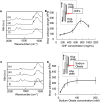
Affinity tests between hydroxyapatite and organic molecules. (A) and (C) FT-IR analyses for the affinity tests of absorbents (A: CNFs and B: oleic acid) on a HAp plate: (i) intact HAp plate; (ii) intact adsorbent; (iii) HAp plate after dropping the adsorbent solution; (iv) HAp plate after washing with sodium chloride solutions. (B) and (D) Shear adhesion strengths of HAp plates bound with different concentrations of (B) CNFs (125, 250, 500, 750 and 1000 mg/ml) and (D) sodium oleate (10, 30, 50, 100 and 200 mM).
Figure 4.
Figure 4.
In vitro fabrication of a 3D trabecular bone-like construct. (A) Schematic design showing the protocol for fabrication of the 3D trabecular bone-like construct. CNFs were mixed and incubated with 20 mM CaCl2 solution for 3 days for fabrication of biomimetic calcospherites, which were centrifugally washed and collected in tubes. The calcospherites were further mixed with CNFs and dropped into a 3D-printed mold. (B) and (C) Digital photographs of the biomimetic calcospherites incubated (B) without and (C) with additional CNFs inside the 3D printed mold. A 3D structure could only be obtained with CNFs to the previously formed biomimetic calcospherites. (D) Secondary electron images of the 3D trabecular bone-like construct showing similar architecture and porosity compared to those of the native trabecular bone. Note that the fusion of calcospherites is observed in the right panel.

Similar articles

References

    1. Robling AG, Castillo AB, Turner CH.. Biomechanical and molecular regulation of bone remodeling. Annu Rev Biomed Eng 2006;8:455–98. - PubMed
    1. Hata K, Takahata Y, Murakami T, Nishimura R.. Transcriptional network controlling endochondral ossification. J Bone Metab 2017;24:75–82. - PMC - PubMed
    1. Shi Y, He G, Lee WC, McKenzie JA, Silva MJ, Long F.. Gli1 identifies osteogenic progenitors for bone formation and fracture repair. Nat Commun 2017;8:2043. - PMC - PubMed
    1. Wang C, Abu-Amer Y, O’Keefe RJ, Shen J.. Loss of Dnmt3b in chondrocytes leads to delayed endochondral ossification and fracture repair. J Bone Miner Res 2018;33:283–97. - PMC - PubMed
    1. Suzuki N, Ohneda O, Minegishi N, Nishikawa M, Ohta T, Takahashi S, Engel JD, Yamamoto M.. Combinatorial Gata2 and Sca1 expression defines hematopoietic stem cells in the bone marrow niche. Proc Natl Acad Sci U S A 2006;103:2202–7. - PMC - PubMed