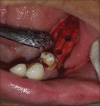
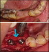
Alveolar ridge split and expansion with simultaneous implant placement in mandibular posterior sites using motorized ridge expanders - modified treatment protocol
- PMID: 36683927
- PMCID: PMC9851358
- DOI: 10.4103/njms.njms_417_21
Alveolar ridge split and expansion with simultaneous implant placement in mandibular posterior sites using motorized ridge expanders - modified treatment protocol
Abstract
Purpose: "The purpose of the study is to evaluate alveolar ridge split and expansion (ARSE) with simultaneous implant placement in mandibular posterior implant sites using motorized ridge expanders."
Background: The ARSE is used in the management of horizontally deficient (narrow) alveolar ridge with optimum bone height available. The ARSE procedure in the posterior mandible has limited application as per literature. The successful cases reported are with extensive procedure of the osteo-mobilization with four corticotomies on buccal side. The authors presented the study of mandibular posterior implant sites using motorized ridge expanders. The ARSE performed here was by only crestal osteotomy simple osteo-condensation and immediate implant insertion.
Materials and methods: The study was prospective type. The sample size was 15 patients and 31 implant sites. The study population included partially edentulous patients between 18 years and 60 years indicated for implant-supported prosthesis. The outcome variables studied included gain in ridge width, cervical bone loss, success of implant, and survival rate. Successful surgical outcome was evaluated by Buser's criteria. The data collected was evaluated by differential statistics.
Conclusion: The minimally invasive technique of one-stage ARSE performed with motorized ridge expander and insertion of implant in the same operative procedure decreases the morbidity, treatment time, number of surgical procedures, and the risk of complications, thereby, increasing patient acceptance. In this study, the authors have used this technique in the posterior mandible for narrow ridges (minimum 3 mm) and obtained promising results. The survival rate of the implants was 100% and the gain in ridge width was 3.2 mm. The author has also recommended the protocol according to bone density of mandible.
Keywords: Alveolar ridge split and expansion; bone density; horizontal ridge deficiency; immediate implant; mandible; motorized ridge expanders; narrow ridge; one stage ridge split; ridge width.
Copyright: © 2022 National Journal of Maxillofacial Surgery.
Conflict of interest statement
There are no conflicts of interest.
Figures


Similar articles
-
Alternative bone expansion technique for implant placement in atrophic edentulous maxilla and mandible.J Oral Implantol. 2011 Aug;37(4):463-71. doi: 10.1563/AAID-JOI-D-10-00028. Epub 2010 Jul 21. J Oral Implantol. 2011. PMID: 20662673 Clinical Trial.
-
Reconstruction of posterior mandibular alveolar ridge deficiencies with the piezoelectric hinge-assisted ridge split technique: a retrospective observational report.J Periodontol. 2010 Nov;81(11):1580-6. doi: 10.1902/jop.2010.100093. Epub 2010 Jul 1. J Periodontol. 2010. PMID: 20594048
-
The effect of modern devices of alveolar ridge split and expansion in the management of horizontally deficient alveolar ridge for dental implant: A systematic review.Natl J Maxillofac Surg. 2023 Sep-Dec;14(3):369-382. doi: 10.4103/njms.njms_423_21. Epub 2022 Jul 22. Natl J Maxillofac Surg. 2023. PMID: 38273919 Free PMC article. Review.
-
Two-Stage Ridge Split at Narrow Alveolar Mandibular Bone Ridges.J Oral Maxillofac Surg. 2017 Oct;75(10):2115.e1-2115.e12. doi: 10.1016/j.joms.2017.05.015. Epub 2017 May 24. J Oral Maxillofac Surg. 2017. PMID: 28623685
-
Interventions for replacing missing teeth: alveolar ridge preservation techniques for dental implant site development.Cochrane Database Syst Rev. 2021 Apr 26;4(4):CD010176. doi: 10.1002/14651858.CD010176.pub3. Cochrane Database Syst Rev. 2021. PMID: 33899930 Free PMC article.
Cited by
-
The efficacy of alveolar ridge split on implants: a systematic review and meta-analysis.BMC Oral Health. 2023 Nov 20;23(1):894. doi: 10.1186/s12903-023-03643-2. BMC Oral Health. 2023. PMID: 37986181 Free PMC article.
-
A Modified Ridge-Splitting Technique to Restore a Completely Edentulous Maxillary Arch With a Cement-Retained Implant Prosthesis.Cureus. 2023 Sep 15;15(9):e45299. doi: 10.7759/cureus.45299. eCollection 2023 Sep. Cureus. 2023. PMID: 37846271 Free PMC article.
-
Utilization of magnetic mallet during dental implantation in narrow mandibular alveolar ridge: A case report.Int J Surg Case Rep. 2025 Jan;126:110679. doi: 10.1016/j.ijscr.2024.110679. Epub 2024 Nov 28. Int J Surg Case Rep. 2025. PMID: 39616743 Free PMC article.
References
-
- Simion M, Baldoni M, Zaffe D. Jawbone enlargement using immediate implant placement associate with a split crest technique and guide tissue regeneration. Int J Periodontics Restorative Dent. 1992;12:462–73. - PubMed
-
- Scipioni A, Bruschi GB, Calesini G. The edentulous ridge expansion technique: A five-year study. Int J Periodontics Restorative Dent. 1994;14:451–9. - PubMed
-
- Sethi A, Kaus T. Maxillary ridge expansion with simultaneous implant placement: 5-year results of an ongoing clinical study. Int J Oral Maxillofac Implants. 2000;15:491–9. - PubMed
-
- Kolerman R, Nissan J, Tal H. Combined osteotome-induced ridge expansion and guided bone regeneration simultaneous with implant placement: A biometric study.Clin Implant Dent Relat Res. 2014;16:691–704. Epub 2013 Jan 25. - PubMed
-
- Bruschi GB, Capparé P, Bravi F, Grande N, Gherlone E, Gastaldi G, et al. Radiographic evaluation of crestal bone level in split-crest and immediate implant placement: Minimum 5-year follow-up. Int J Oral Maxillofac Implants. 2017;32:114–20. - PubMed
LinkOut - more resources
Full Text Sources
