. 2022 May 4;8(1):e10322.
doi: 10.1002/btm2.10322. eCollection 2023 Jan.

Sound-guided assessment and localization of pulmonary air leak

Affiliations

Sound-guided assessment and localization of pulmonary air leak

Meghan R Pinezich et al. Bioeng Transl Med. .

Abstract

Pulmonary air leak is the most common complication of lung surgery, with air leaks that persist longer than 5 days representing a major source of post-surgery morbidity. Clinical management of air leaks is challenging due to limited methods to precisely locate and assess leaks. Here, we present a sound-guided methodology that enables rapid quantitative assessment and precise localization of air leaks by analyzing the distinct sounds generated as the air escapes through defective lung tissue. Air leaks often present after lung surgery due to loss of tissue integrity at or near a staple line. Accordingly, we investigated air leak sounds from a focal pleural defect in a rat model and from a staple line failure in a clinically relevant swine model to demonstrate the high sensitivity and translational potential of this approach. In rat and swine models of free-flowing air leak under positive pressure ventilation with intrapleural microphone 1 cm from the lung surface, we identified that: (a) pulmonary air leaks generate sounds that contain distinct harmonic series, (b) acoustic characteristics of air leak sounds can be used to classify leak severity, and (c) precise location of the air leak can be determined with high resolution (within 1 cm) by mapping the sound loudness level across the lung surface. Our findings suggest that sound-guided assessment and localization of pulmonary air leaks could serve as a diagnostic tool to inform air leak detection and treatment strategies during video-assisted thoracoscopic surgery (VATS) or thoracotomy procedures.

Keywords: Lung disease; digital medicine; lung volume reduction surgery; minimally‐invasive diagnosis; sound analysis.


Conflict of interest statement

No competing interests to disclose.

Figures

FIGURE 1
FIGURE 1
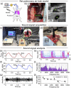
Assessment and localization of pulmonary air leak using sound analysis. (a) Schematic of pulmonary air leak with computer‐assisted sound analysis system. Mic: microphone. F: frequency (b) Process flow diagram of air leak sound analysis for severity prediction and localization to improve clinical decision‐making for patients with air leak
FIGURE 2
FIGURE 2
Analysis of pulmonary air leak sounds in rat lungs. (a) Schematic of needle puncture and air leak induction in rat lung. S: pressure and flow sensors. (b) (i) Puncture wound in rat lung. (ii) Air leak was induced by puncturing the lung with a needle (18‐gauge or 16‐gauge). (iii) Focal puncture wound (diameter: 1.3 mm). (c) Photograph of setup to monitor pressure and record air leak sounds in rat lungs. (d) Photograph of microphone positioned above the punctured rat lung for sound recording. Mic: microphone. (e) Pressure of inhaled air (P airway) measured at the trachea during sound recording. Insp: inspiration. Exp: expiration. (f) A‐weighted sound pressure level (SPL). (g) Normalized amplitude of the acquired sound signal. (h) Spectrogram of the air leak sound calculated from the recorded sound showing sound frequency distribution and density. Freq: frequency. (i) Representative air leak power spectra obtained via Fourier transform. (j) Inverse relationship between band power and frequency in air leak power spectra (P < 0.001, R 2 = 0.702). Y = −0.00231X – 49.1, where X is frequency and Y is frequency band power
FIGURE 3
FIGURE 3
Analysis of pulmonary air leak sounds in swine lung staple line failure model. (a) Schematic of swine wedge resection and staple line failure model to induce pulmonary air leak for sound analysis in situ. S: pressure and flow sensors. (b) Photograph of right middle lobe with air bubbling at site of staple line failure air leak. (c) Radiograph of radiopaque dye leaking from the lung parenchyma at site of staple line failure air leak. (d) Photograph of air leak sound acquisition in situ. Mic: microphone. (e) Pressure of inhaled air (P airway) measured at the trachea during sound recording. Insp: inspiration. Exp: expiration. (f) Normalized amplitude. (g) A‐weighted sound pressure level (SPL). (h) Spectrogram of the air leak sound calculated from the recorded sound showing sound frequency distribution and density. Freq: frequency. (i) Spectrogram showing sound frequency distribution and density of mild air leak. Freq: frequency. V Loss: tidal volume loss. Br. breath (j) Power spectra of air leaks of varying severity. (k) loudness of air leak sounds of varying severity
FIGURE 4
FIGURE 4
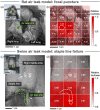
Localization of air leak site by measurement of relative loudness. (a) Rat lung photograph and (b) corresponding breath sound intensity matrix heat map. (c) Swine lung photograph and (d) corresponding breath sound intensity matrix heat map. The value in each array indicates the measured loudness normalized to the maximum loudness measured at the air leak site (dotted regions). Inset: overview photographs of analyzed regions of lung

Similar articles

Cited by

  • Lung-Mimetic Hydrofoam Sealant to Treat Pulmonary Air Leak.
    Pinezich MR, Mir M, Graney PL, Tavakol DN, Chen J, Hudock MR, Gavaudan O, Chen P, Kaslow SR, Reimer JA, Van Hassel J, Guenthart BA, O'Neill JD, Bacchetta M, Kim J, Vunjak-Novakovic G. Pinezich MR, et al. Adv Healthc Mater. 2024 May;13(13):e2303026. doi: 10.1002/adhm.202303026. Epub 2024 Feb 14. Adv Healthc Mater. 2024. PMID: 38279961 Free PMC article.

References

    1. Okereke I, Murthy SC, Alster JM, Blackstone EH, Rice TW. Characterization and importance of air leak after lobectomy. Ann Thorac Surg. 2005;79(4):1167‐1173. doi:10.1016/j.athoracsur.2004.08.069 - DOI - PubMed
    1. Marshall MB, Deeb ME, Bleier JIS, et al. Suction vs water seal after pulmonary resection: a randomized prospective study. Chest. 2002;121(3):831‐835. doi:10.1378/chest.121.3.831 - DOI - PubMed
    1. Baringer K, Talbert S. Chest drainage systems and management of air leaks after a pulmonary resection. J Thorac Dis. 2017;9(12):5399‐5403. doi:10.21037/jtd.2017.11.15 - DOI - PMC - PubMed
    1. Yano M, Iwata H, Hashizume M, et al. Adverse events of lung tissue stapling in thoracic surgery. Ann Thorac Cardiovasc Surg. 2014;20(5):370‐377. doi:10.5761/atcs.oa.13-00161 - DOI - PubMed
    1. Eckert CE, Harris JL, Wong JB, et al. Preclinical quantification of air leaks in a physiologic lung model: effects of ventilation modality and staple design. Med Devices (Auckl). 2018;11:433‐442. doi:10.2147/MDER.S184851 - DOI - PMC - PubMed

LinkOut - more resources