. 2022 Jul 1;8(1):e10366.
doi: 10.1002/btm2.10366. eCollection 2023 Jan.

Cold air plasma improving rheumatoid arthritis via mitochondrial apoptosis pathway

Affiliations

Cold air plasma improving rheumatoid arthritis via mitochondrial apoptosis pathway

Chengbiao Ding et al. Bioeng Transl Med. .

Abstract

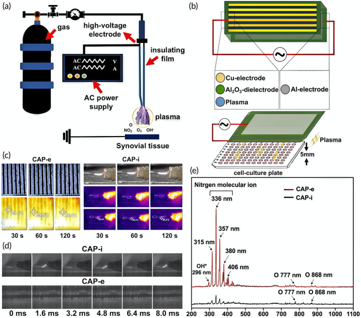
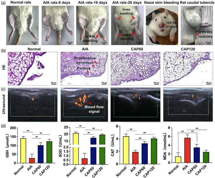
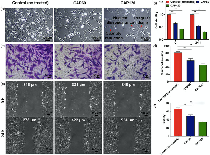
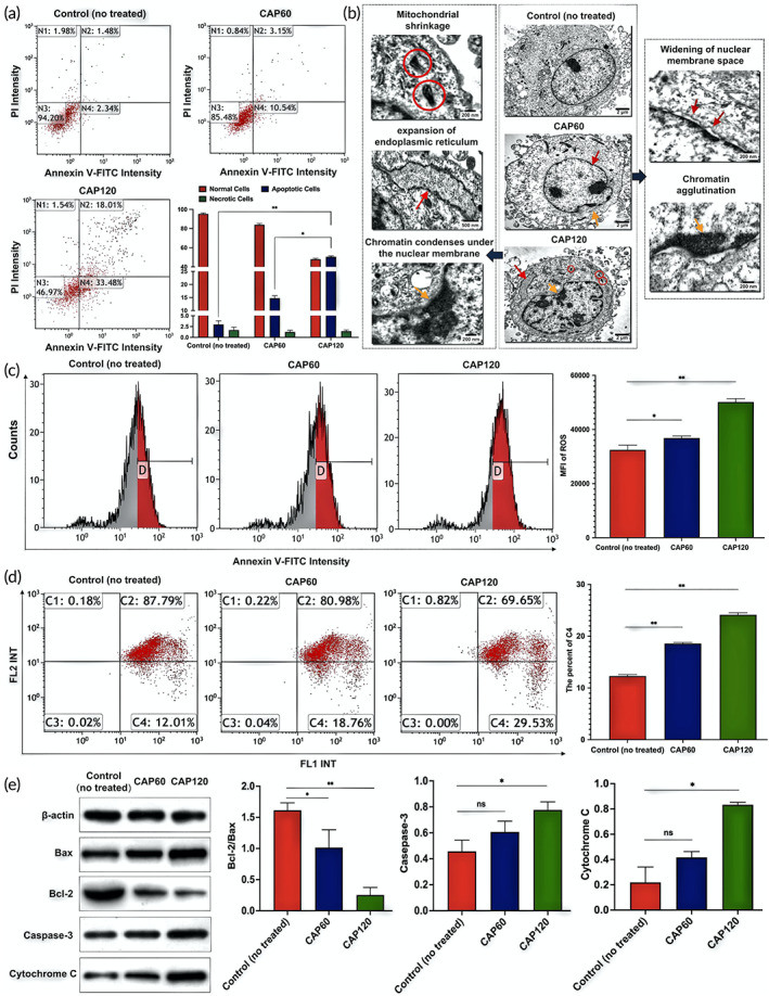
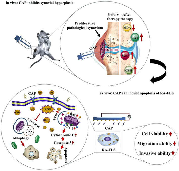
Rheumatoid arthritis (RA) has plagued physicians and patients for years due to the lack of targeted treatment. In this study, inspired by the commonality between rheumatoid arthritis fibroblast-like synoviocytes (RA-FLS) and cancer cells, the therapeutic effects of cold air plasma (CAP) on RA are studied systematically and thoroughly. In/ex vivo results show that CAP with the proper dosage significantly relieves symptoms including synovial hyperplasia, inflammatory infiltration, and angiogenesis and eliminates the root cause by triggering the self-antioxidant capability of the surrounding tissue. The mechanism on the molecular and cellular level is also revealed that the spontaneous reactive oxygen species (ROS) cascade induces the mitochondrial apoptosis pathway on RA-FLS. This study reveals a new strategy for targeted treatment of RA and the mechanistic study provides the theoretical foundation for future development of plasma medicine.

Keywords: ROS; cold air plasma; mitochondrial apoptosis pathway; rheumatoid arthritis.


Conflict of interest statement

The authors declare no competing interests.

Figures

FIGURE 1
FIGURE 1
Illustration of cold air plasma (CAP) devices and physical characteristics of plasma. (a) Schematic diagram of CAP‐i device. (b) Schematic diagram of CAP‐e device. (c) The infrared thermometer measures the working temperature of CAP‐i and CAP‐e device at different times. (d) The discharge image was recorded by means of a high‐speed camera device (phantom) with 600 frames per second. (e) Emission spectra of CAP‐i and CAP‐e between 200 and 1100 nm
FIGURE 2
FIGURE 2
Cold air plasma (CAP) in vivo treatment can reduce the inflammation and proliferation of synovium in adjuvant‐induced arthritis (AIA) rats, inhibit blood flow and improve oxidative stress indicators. (a) Establishment of AIA rat model. (b) Changes in HE staining of the synovial tissue of the knee joint in each group of rats. (c) Changes in ultrasound blood flow in the synovial membrane of the knee joint in each group of rats. (d) Changes of oxidative stress indexes before and after treatment, ** indicates statistical differences: **p < 0.01
FIGURE 3
FIGURE 3
Cold air plasma (CAP) treatment ex vivo can reduce the cell viability, invasion, and migration of rheumatoid arthritis fibroblast‐like synoviocytes (RA‐FLS). (a) Cell morphology was observed by inverted microscope (magnification: 100×) at different time points of CAP treatment. (b) CCK‐8 assay of cell viability in each group. ** indicates statistical differences: **p < 0.01. (c) Experimental pictures of RA‐FLS invasion in each group. (d) Experimental pictures of RA‐FLS invasion in each group. (e) Pictures of RA‐FLS migration distance at 0 and 24 h after CAP intervention in each group. (f) Statistics of cell migration rate in each group. ** indicates statistical differences: **p < 0.01.
FIGURE 4
FIGURE 4
Changes of apoptosis, morphology, intracellular reactive oxygen species (ROS) content, mitochondrial membrane potential (MMP) and related apoptotic proteins in rheumatoid arthritis fibroblast‐like synoviocytes (RA‐FLS) before and after cold air plasma (CAP) intervention ex vivo: (a) Effect of CAP on apoptosis of RA‐FLS cells. Include the ratios of necrotic, apoptotic, and viable cells of each group within 24 h after CAP intervention and the apoptosis indexes of RA‐FLS cells of each group. * and ** indicates a significant difference between the groups, equal to *p < 0.05 and **p < 0.01. (b) Cell morphology at different time of the CAP treatment observed by transmission electron microscopy. (c) ROS content and statistical difference in the control group (untreated), the CAP60 group, and the CAP120 group, ** indicates a significant difference between the groups (p < 0.01). (d) JC‐1 fluorescence intensity in each group, C2 represents the proportion of cells in good condition, and C4 represents the proportion of cells with reduced membrane potential, * and ** indicates a significant difference between the groups, equal to *p < 0.05 and **p < 0.01. (e) Western blot detected the expressions of Bcl‐2, Bax, caspase‐3 and cytochrome c in RA‐FLS cells; Bcl‐2/Bax ratio; Relative expression of caspase‐3 protein; relative expression of cytochrome c protein. * and ** indicate that there are significant differences between the two groups, which are equal to *p < 0.05 and **p < 0.01 respectively. “ns” indicates that there is no difference between the two groups, which is equal to ns p > 0.05. Before and after CAP intervention, there was no significant difference in the protein content of activated caspase‐3 between CAP60 group and control (no treated) group (p = 0.095), and there was no significant difference in the protein content of cytochrome c between CAP60 group and control (no treated) group (p = 0.131).
FIGURE 5
FIGURE 5
Schematic diagram of the mechanism of CAP's therapeutic effect on AIA rat joint synovium in vivo and ex vivo

References

    1. Liang C, Li J, Lu C, et al. HIF1α inhibition facilitates Leflunomide‐AHR‐CRP signaling to attenuate bone erosion in CRP‐aberrant rheumatoid arthritis. Nat Commun. 2019;10:4579. - PMC - PubMed
    1. Yang S, Zhang X, Chen J, et al. Induced, but not natural, regulatory T cells retain phenotype and function following exposure to inflamed synovial fibroblasts. Sci. Adv. 2020;6(44):eabb0606. - PMC - PubMed
    1. Orr C, Vieira‐Sousa E, Boyle DL, et al. Synovial tissue research: a state‐of‐the‐art review. Nat Rev Rheumatol. 2017;13:463‐475. - PubMed
    1. Machado GC, Abdel‐Shaheed C, Underwood M, Day RO. Non‐steroidal anti‐inflammatory drugs (NSAIDs) for musculoskeletal pain. BMJ. 2021;372:n104. - PubMed
    1. Johnson S, Katyal N, Narula N, Govindarajan R. Adverse side effects associated with corticosteroid therapy: a study in 39 pPatients with generalized Myasthenia Gravis. Med Sci Monit. 2021;27:e933296. - PMC - PubMed

LinkOut - more resources