. 2022 Jul 12;8(1):e10368.
doi: 10.1002/btm2.10368. eCollection 2023 Jan.

Photothermally responsive theranostic nanocomposites for near-infrared light triggered drug release and enhanced synergism of photothermo-chemotherapy for gastric cancer

Affiliations

Photothermally responsive theranostic nanocomposites for near-infrared light triggered drug release and enhanced synergism of photothermo-chemotherapy for gastric cancer

Taicheng Zhou et al. Bioeng Transl Med. .

Abstract

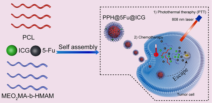
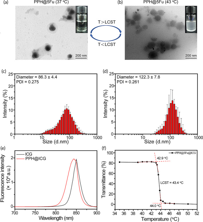
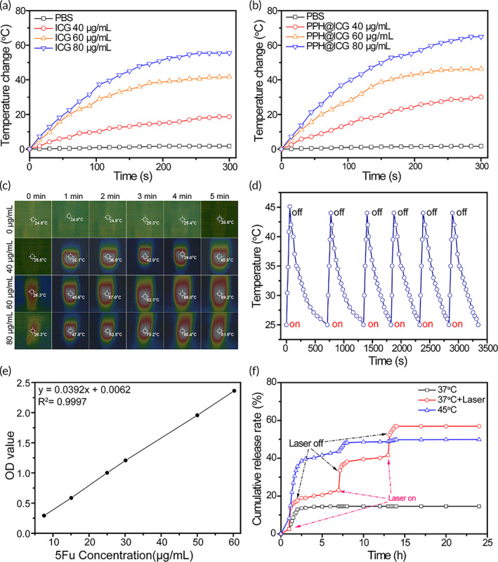
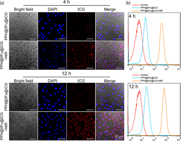
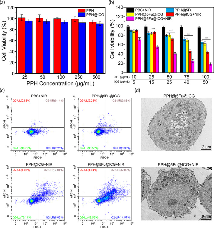
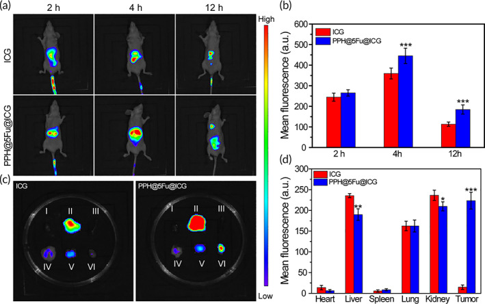
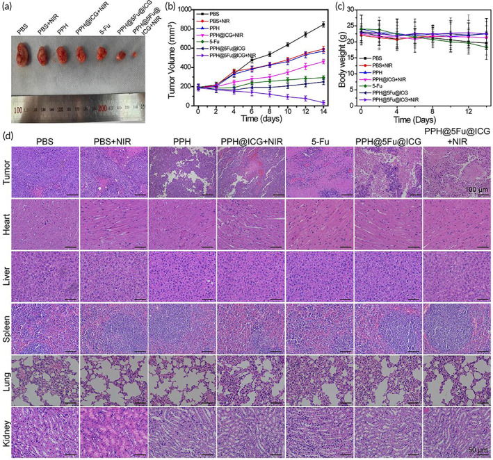
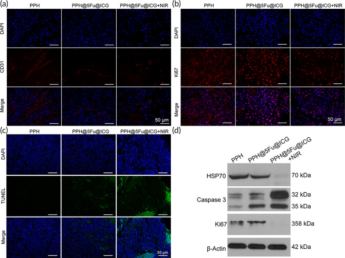
Near-infrared (NIR) photothermal therapy plays a critical role in the cancer treatment and diagnosis as a promising carcinoma treatment modalities nowadays. However, development of clinical application has been greatly limited due to the inefficient drug release and low tumor accumulation. Herein, we designed a NIR-light triggered indocyanine green (ICG)-based PCL core/P(MEO2MA-b-HMAM) shell nanocomposites (PPH@ICG) and evaluated their therapeutic effects in vitro and in vivo. The anticancer drug 5-fluorouracil (5Fu) and the photothermal agent ICG were loaded into a thermo-sensitive micelle (PPH@5Fu@ICG) by self-assembly. The nanoparticles formed were characterized using transmission electron microscopy, dynamic light scattering, and fluorescence spectra. The thermo-sensitive copolymer (PPH@5Fu@ICG) showed a great temperature-controlled drug release response with lower critical solution temperature. In vitro cellular uptake and TEM imaging proved that PPH@5Fu@ICG nanoparticles can home into the lysosomal compartments under NIR. Moreover, in gastric tumor-bearing nude mice, PPH@5Fu@ICG + NIR group exhibited excellent improvement in antitumor efficacy based on the NIR-triggered thermo-chemotherapy synergy, both in vitro and in vivo. In summary, the proposed strategy of synergistic photo-hyperthermia chemotherapy effectively reduced the 5Fu dose, toxic or side effect, which could serve as a secure and efficient approach for cancer theranostics.

Keywords: chemotherapy; gastric cancer; nanocarriers; photothermal therapy; photothermally responsive.


Conflict of interest statement

There are no conflicts to declare.

Figures

SCHEME 1
SCHEME 1
Schematic illustration for the preparation of PPH@5Fu@ICG and its related mechanisms for the treatment of GC
FIGURE 1
FIGURE 1
Characterization of the temperature‐sensitive PPH@5Fu nanoparticles. (a) TEM images of PPH@5Fu nanoparticles at 37°C. (b) TEM images of PPH@5Fu nanoparticles at 43°C. (c) Size distribution of PPH@5Fu nanoparticles at 37°C. (d) Size distribution of PPH@5Fu nanoparticles at 43°C. (e) The fluorescence emission spectra of ICG and PPH@ICG in the wavelength from 700 to 900 nm. (f) The transmittance curves of PPH@5Fu@ICG micelles
FIGURE 2
FIGURE 2
Photothermal properties of the nanoparticles. Photothermal heating curves of free ICG (a) and PPH@ICG (b) with various concentrations under continuous laser irradiation (808 nm, 1 W/cm2). (c) Infrared thermographs of PPH@ICG under continuous laser irradiation (808 nm, 1 W/cm2) for various irradiation times. (d) Photothermal stability evaluation of PPH@ICG with laser switch‐on and switch‐off treatment for six cycles. (e) The standard curve of 5Fu. (f) In vitro cumulative 5Fu release rate of PPH@ICG@5Fu at 37°C (with or without of laser irradiation) and 45°C. 5Fu, 5‐fluorouracil; ICG, indocyanine green
FIGURE 3
FIGURE 3
In vitro cellular uptake. (a) Confocal laser images of AGS cells after incubation with PPH@5Fu@ICG and PPH@5Fu@ICG + NIR for 4 and 12 h. (b) Endocytosis of PPH@5Fu@ICG and PPH@5Fu@ICG + NIR by AGS cells at 4 and 12 h post‐incubation via flow cytometry. Experiments were repeated three times. 5Fu, 5‐fluorouracil
FIGURE 4
FIGURE 4
Enhanced cytotoxicity by NIR laser‐driven drug release. (a) Cell viability of AGS cells treated with PPH and PPH@ICG at various ICG concentrations. (b) Cell viability of AGS cells treated with PBS + NIR, PPH@5Fu, PPH@5Fu@ICG, PPH@ICG + NIR, and PPH@5Fu@ICG + NIR at various 5Fu or ICG concentrations. (c) Flow cytometry analysis of AGS cells after incubation with PBS + NIR, PPH@5Fu@ICG, PPH@ICG + NIR, and PPH@5Fu@ICG + NIR. Double stained cells were considered as late apoptotic/necrotic cells. (d) TEM images of AGS cells after incubation with PPH@5Fu@ICG and PPH@5Fu@ICG + NIR for 12 h. Experiments were repeated three times. 5Fu, 5‐fluorouracil; ICG, indocyanine green; NIR, near‐infrared
FIGURE 5
FIGURE 5
In vivo NIR fluorescence imaging AGS tumor‐bearing nude mice. (a) In vivo NIR fluorescence images of AGS tumor‐bearing nude mice 12 h receiving intravenous injection of ICG or PPH@5Fu@ICG. (b) Quantitation of the ICG fluorescence intensity of mice at different time points. (c) NIR fluorescence images of the isolated major organs and tumors 12 h post‐injection with ICG or PPH@5Fu@ICG (I, heart; II, liver; III, spleen; IV, kidney; V, Lung; VI, tumor). (d) Quantitation of ICG fluorescence intensity of individual organs and tumor. Experiments were repeated three times. 5Fu, 5‐fluorouracil; ICG, indocyanine green; NIR, near‐infrared
FIGURE 6
FIGURE 6
In vivo antitumor performance. (a) Photographs of representative tumors resected from different groups at 14 days. (b) Tumor growth curve in terms of volume. (c) Body weight of mice in different groups after treatments. (d) H&E staining images of tumor and major organs (heart, liver, spleen, lung, and kidney) slices after various treatments. Experiments were repeated three times
FIGURE 7
FIGURE 7
(a) Immunofluorescence of CD31 in the tumors. (b) Immunofluorescence of Ki67 in the tumors. (c) Immunofluorescence of TUNEL in the tumors. (d) WB analysis of HSP70, Caspase‐3, and Ki67. Experiments were repeated three times. HSP70, heat shock protein 70; WB, western blotting

References

    1. Chen D, Liu Z, Liu W, et al. Predicting postoperative peritoneal metastasis in gastric cancer with serosal invasion using a collagen nomogram. Nat Commun. 2021;12:179. - PMC - PubMed
    1. El‐Rayes BF, Zalupski M, Bekai‐Saab T, et al. A phase II study of bevacizumab, oxaliplatin, and docetaxel in locally advanced and metastatic gastric and gastroesophageal junction cancers. Ann Oncol. 2010;21:1999‐2004. - PMC - PubMed
    1. Li X, Pasche B, Zhang W, Chen K. Association of MUC16 mutation with tumor mutation load and outcomes in patients with gastric cancer. JAMA Oncol. 2018;4:1691‐1698. - PMC - PubMed
    1. Yuan L, Xu ZY, Ruan SM, Mo S, Qin JJ, Cheng XD. Long non‐coding RNAs towards precision medicine in gastric cancer: early diagnosis, treatment, and drug resistance. Mol Cancer. 2020;19:96. - PMC - PubMed
    1. Lu J, Liu X, Liao YP, et al. Nano‐enabled pancreas cancer immunotherapy using immunogenic cell death and reversing immunosuppression. Nat Commun. 2017;8:1811. - PMC - PubMed

LinkOut - more resources