

Single-cell transcriptomes and T cell receptors of vaccine-expanded apolipoprotein B-specific T cells
- PMID: 36684560
- PMCID: PMC9849899
- DOI: 10.3389/fcvm.2022.1076808
Single-cell transcriptomes and T cell receptors of vaccine-expanded apolipoprotein B-specific T cells
Abstract
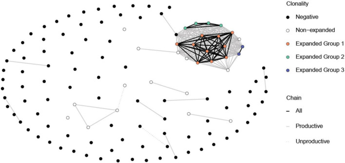
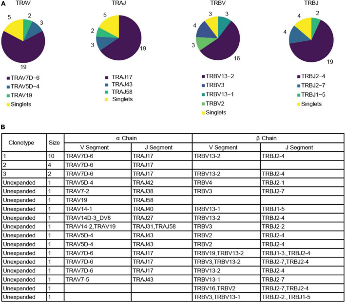
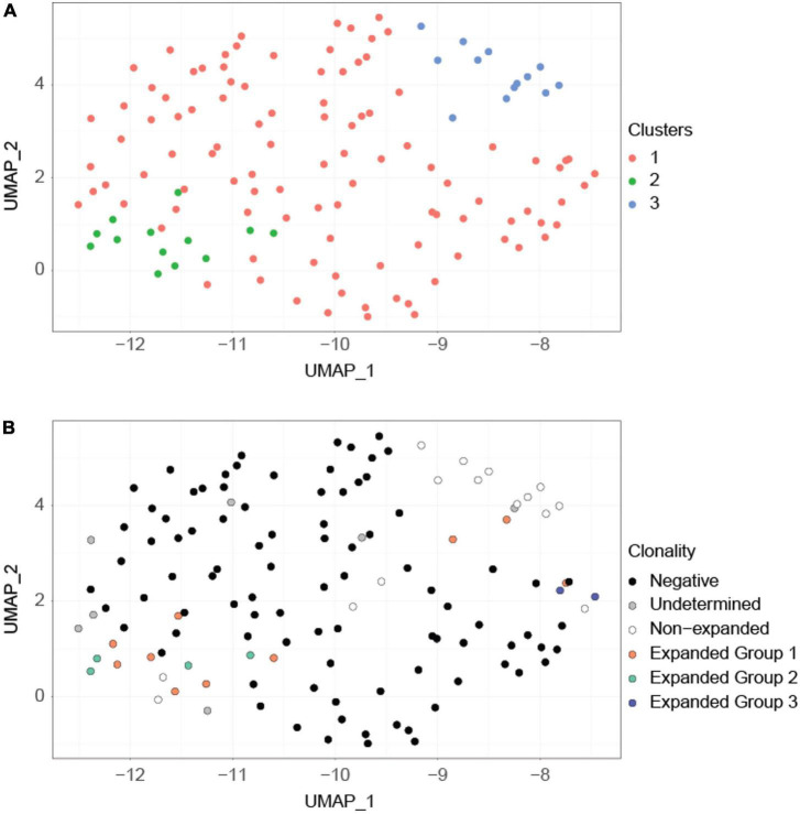
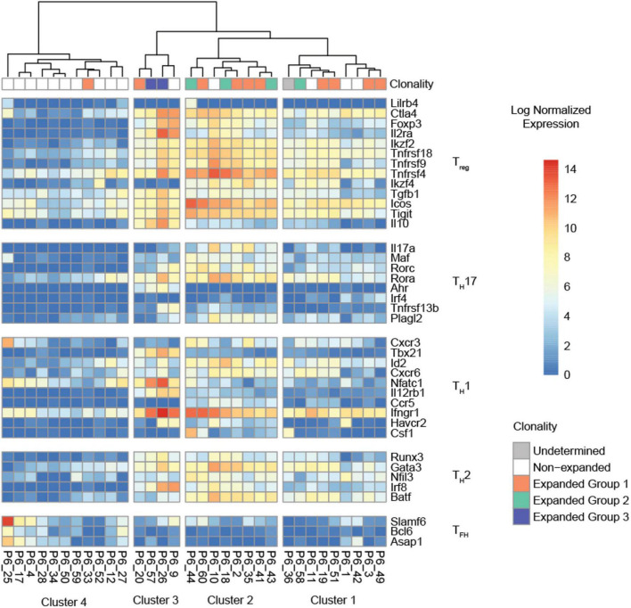
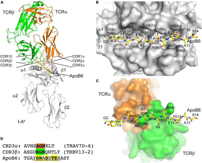
Atherosclerotic cardiovascular diseases are the major cause of death worldwide. CD4 T cells responding to Apolipoprotein B (ApoB), the core protein of most lipoproteins, have been identified as critical disease modulators. In healthy individuals, ApoB-reactive (ApoB+) CD4 T cells are mostly regulatory T cells (Tregs), which exert anti-inflammatory effects. Yet, they may obtain pro-inflammatory features and thus become proatherogenic. Evidence from animal studies suggests that vaccination against certain major histocompatibility complex (MHC) II-binding ApoB peptides induces an expansion of ApoB+ Tregs and thus confers atheroprotection. To date, in-depth phenotyping of vaccine-expanded ApoB+ T cells has not yet been performed. To this end, we vaccinated C57BL/6J mice with the ApoB-peptide P6 (ApoB978-993 TGAYSNASSTESASY) and performed single-cell RNA sequencing of tetramer-sorted P6+ T cells. P6+ cells were clonally expanded (one major, two minor clones) and formed a transcriptional cluster distinct from clusters mainly containing non-expanded P6+ and P6- cells. Transcriptomic profiling revealed that most expanded P6+ cells had a strong Treg signature and highly expressed genes mediating suppressive functions. Yet, some expanded P6+ cells only had a residual Treg signature and expressed genes related to T helper 1 (TH1) cells, which are proatherogenic. Modeling the T cell receptor (TCR) and P6:MHC-II interaction showed that only three amino acid residues in the α and β chain contact the P6 peptide in the MHC-II groove and thus determine the specificity of this TCR to P6. Our data begin to reveal the vaccination-induced response to an ApoB epitope.
Keywords: ApoB; P6; atherosclerosis vaccine; regulatory T cells; single-cell RNA-sequencing.
Copyright © 2023 Nettersheim, Ghosheh, Winkels, Kobiyama, Durant, Armstrong, Brunel, Roy, Dileepan, Jenkins, Zajonc and Ley.
Conflict of interest statement
KL was founder and co-owner of Atherovax, Inc. He received no compensation from Atherovax. No Atherovax funds were used in this study. The remaining authors declare that the research was conducted in the absence of any commercial or financial relationships that could be construed as a potential conflict of interest.
Figures
Similar articles
-
Pathogenic Autoimmunity in Atherosclerosis Evolves From Initially Protective Apolipoprotein B100-Reactive CD4+ T-Regulatory Cells.Circulation. 2020 Sep 29;142(13):1279-1293. doi: 10.1161/CIRCULATIONAHA.119.042863. Epub 2020 Jul 24. Circulation. 2020. PMID: 32703007 Free PMC article.
-
Atheroprotective vaccination with MHC-II-restricted ApoB peptides induces peritoneal IL-10-producing CD4 T cells.Am J Physiol Heart Circ Physiol. 2017 Apr 1;312(4):H781-H790. doi: 10.1152/ajpheart.00798.2016. Epub 2017 Jan 13. Am J Physiol Heart Circ Physiol. 2017. PMID: 28087520 Free PMC article.
-
Autoimmune Regulator (AIRE) Deficiency Does Not Affect Atherosclerosis and CD4 T Cell Immune Tolerance to Apolipoprotein B.Front Cardiovasc Med. 2022 Jan 13;8:812769. doi: 10.3389/fcvm.2021.812769. eCollection 2021. Front Cardiovasc Med. 2022. PMID: 35097028 Free PMC article.
-
ApoB-Specific CD4+ T Cells in Mouse and Human Atherosclerosis.Cells. 2021 Feb 19;10(2):446. doi: 10.3390/cells10020446. Cells. 2021. PMID: 33669769 Free PMC article. Review.
-
Biological drug and drug delivery-mediated immunotherapy.Acta Pharm Sin B. 2021 Apr;11(4):941-960. doi: 10.1016/j.apsb.2020.12.018. Epub 2020 Dec 31. Acta Pharm Sin B. 2021. PMID: 33996408 Free PMC article. Review.
Cited by
-
Atherosclerosis antigens as targets for immunotherapy.Nat Cardiovasc Res. 2023 Dec;2(12):1129-1147. doi: 10.1038/s44161-023-00376-x. Epub 2023 Dec 11. Nat Cardiovasc Res. 2023. PMID: 39196152 Review.
-
Mini-Review: Immunogenic epitopes in apolipoprotein B-100 for atheroprotective immunization.Front Cardiovasc Med. 2024 Aug 15;11:1448664. doi: 10.3389/fcvm.2024.1448664. eCollection 2024. Front Cardiovasc Med. 2024. PMID: 39211769 Free PMC article. Review.
-
Late-rising CD4 T cells resolve mouse cytomegalovirus persistent replication in the salivary gland.PLoS Pathog. 2024 Jan 18;20(1):e1011852. doi: 10.1371/journal.ppat.1011852. eCollection 2024 Jan. PLoS Pathog. 2024. PMID: 38236791 Free PMC article.
-
PD-1 and CD73 on naive CD4+ T cells synergistically limit responses to self.Nat Immunol. 2025 Jan;26(1):105-115. doi: 10.1038/s41590-024-02021-6. Epub 2024 Nov 21. Nat Immunol. 2025. PMID: 39572641 Free PMC article.
References
Grants and funding
LinkOut - more resources
Full Text Sources
Molecular Biology Databases
Research Materials
Miscellaneous