

Whole-cycle analysis of echocardiographic tissue Doppler velocities as a marker of biological age
- PMID: 36684568
- PMCID: PMC9846028
- DOI: 10.3389/fcvm.2022.1040647
Whole-cycle analysis of echocardiographic tissue Doppler velocities as a marker of biological age
Abstract
Purpose: Tissue Doppler imaging (TDI) is a sensitive marker of impaired cardiac function and different phases of the TDI curve carry different prognostic information. It is not known how continuous TDI curves change with age in normal subjects, and whether these changes differ from changes seen in individuals at risk of future cardiac events.
Methods: A total of 1,763 individuals from the general population were examined with color TDI at the septal and lateral mitral sites. A low-risk group was defined as without cardiac risk factors (hypertension, diabetes or ischemic heart disease) at baseline and without any cardiac events (cardiovascular death or admission due to either heart failure or acute myocardial infarction) during 10-years follow-up. All TDI curves were corrected for heart rate, and whole-cycle analysis of age-related changes to TDI velocities was performed in both low-risk (n = 881) and high-risk individuals (n = 882).
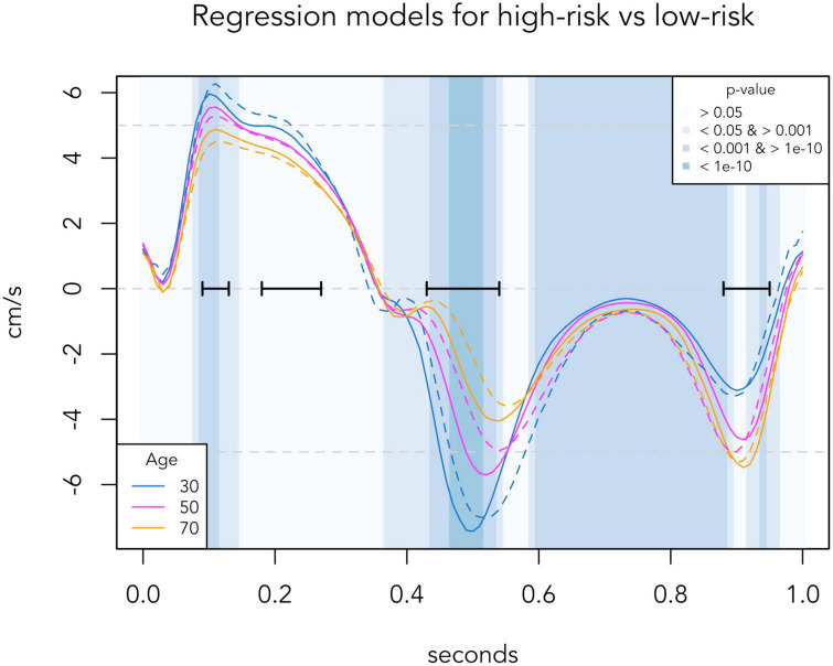
Results: In the low-risk population, four phases where myocardial velocity differed most (p < 10-10) according to age were identified [in a standardized cardiac cycle of 1 second (s)]: Systolic peak (0.09-0.13 s), systolic plateau (0.18-0.27 s), early diastole (0.43-0.54 s) and late diastole (0.88-0.95 s). With increasing age, systolic velocities decreased, early diastolic velocities decreased and had delayed peak, and late diastolic velocities increased until age 70 and then stopped increasing. In the high-risk population, comparison to corresponding age groups of the low-risk population showed: Lower early diastolic velocities in 20-40-year-olds; higher late diastolic velocities and lower peak systolic velocities in 40-60-year-olds; further decreased systolic velocities including the systolic plateau and decreased late diastolic velocities in 60-year-olds. The time segments around the systolic peak (p = 0.002) and early diastole (p < 0.001) differed significantly between the high-risk and low-risk population, thus making it possible to use the individual age gap between a TDI-derived biological age and the real chronological age as a tool to discriminate high-risk individuals from low-risk individuals.
Conclusion: We found that individuals with cardiac risk factors display findings compatible with an accelerated aging of the heart and thus propose TDI-derived biological age as a tool to identify high-risk patients.
Keywords: accelerated aging; biological age; cardiac degeneration; healthy aging; tissue Doppler imaging.
Copyright © 2023 Wang, Olsen, Taraldsen and Mogelvang.
Conflict of interest statement
The authors declare that the research was conducted in the absence of any commercial or financial relationships that could be construed as a potential conflict of interest.
Figures


Similar articles
-
Tissue Doppler imaging adds incremental value in predicting exercise capacity in patients with congestive heart failure.Heart Vessels. 2007 Jul;22(4):237-44. doi: 10.1007/s00380-006-0961-x. Epub 2007 Jul 20. Heart Vessels. 2007. PMID: 17653517
-
Haemodialysis: effects of acute decrease in preload on tissue Doppler imaging indices of systolic and diastolic function of the left and right ventricles.Eur J Echocardiogr. 2008 Jul;9(4):530-5. doi: 10.1093/ejechocard/jen125. Epub 2008 Mar 27. Eur J Echocardiogr. 2008. PMID: 18490307
-
Load-independent parameters of diastolic and systolic function by speckle tracking and tissue doppler in hemodialysis patients.Rev Port Cardiol. 2008 Sep;27(9):1011-25. Rev Port Cardiol. 2008. PMID: 19044173 English, Portuguese.
-
Peak early diastolic mitral annulus velocity by tissue Doppler imaging adds independent and incremental prognostic value.J Am Coll Cardiol. 2003 Mar 5;41(5):820-6. doi: 10.1016/s0735-1097(02)02921-2. J Am Coll Cardiol. 2003. PMID: 12628728
-
Clinical aspects of left ventricular diastolic function assessed by Doppler echocardiography following acute myocardial infarction.Dan Med Bull. 2001 Nov;48(4):199-210. Dan Med Bull. 2001. PMID: 11767125 Review.
Cited by
-
EchoAGE: Echocardiography-based Neural Network Model Forecasting Heart Biological Age.Aging Dis. 2024 Jul 28;16(4):2383-2397. doi: 10.14336/AD.2024.0615. Aging Dis. 2024. PMID: 39226165 Free PMC article.
-
Current and Clinically Relevant Echocardiographic Parameters to Analyze Left Atrial Function.J Cardiovasc Dev Dis. 2024 Aug 5;11(8):241. doi: 10.3390/jcdd11080241. J Cardiovasc Dev Dis. 2024. PMID: 39195149 Free PMC article. Review.
-
A biomarker framework for cardiac aging: the Aging Biomarker Consortium consensus statement.Life Med. 2023 Sep 27;2(5):lnad035. doi: 10.1093/lifemedi/lnad035. eCollection 2023 Oct. Life Med. 2023. PMID: 39872891 Free PMC article.
-
Impact of Left Atrial Ablation on the Atrial Contractile Function: Insights From Intracardiac Echocardiography and Electroanatomical Mapping in Persistent Atrial Fibrillation Ablation.J Arrhythm. 2025 Aug 21;41(4):e70179. doi: 10.1002/joa3.70179. eCollection 2025 Aug. J Arrhythm. 2025. PMID: 40861255 Free PMC article.
References
-
- Nagueh S, Smiseth O, Appleton C, Byrd B, Dokainish H, Edvardsen T, et al. Recommendations for the evaluation of left ventricular diastolic function by echocardiography: an update from the American Society of echocardiography and the european association of cardiovascular imaging. Eur Heart J Cardiovasc Imaging. (2016) 17:1321–60. 10.1093/ehjci/jew082 - DOI - PubMed
LinkOut - more resources
Full Text Sources
Miscellaneous