

A rare case of brain metastatic of primary mediastinal yolk sac tumor
- PMID: 36684631
- PMCID: PMC9849995
- DOI: 10.1016/j.radcr.2022.12.028
A rare case of brain metastatic of primary mediastinal yolk sac tumor
Abstract
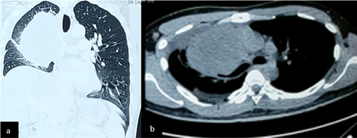
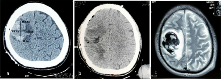
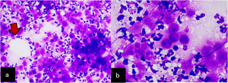
Primary yolk sac tumors are extragonadal germ cell tumors commonly seen in children and young adults. They are more common in men. Germ cells tumor on histopathological characteristics is classified as seminoma and non-seminomatous (NSGC). The rarest form of NSGC is an extragonadal yolk sac tumor of mediastinum. Clinical presentations are not specific and may imitate other chronic disease such as other malignancies or tuberculosis such as chest discomfort, vena cava superior syndrome, fever, weight loss, and chronic cough. Immunohistochemistry showed a positive result in Alpha-fetoprotein and pan-cytokeratin. Due to its rarity, brain metastases' clinical signs and symptoms, anatomical sites, and characteristics are less well documented. However, the metastatic brain process gave similar histological findings to the primary site. Additional radiological and laboratory tests can be carried out to identify other metastatic processes. Standardized treatment of primary mediastinal sac tumors with brain metastasis has not yet been established. Combining chemotherapy, surgery and radiation treatment could improve overall outcomes and prognosis. We present a scarce case of primary mediastinal yolk sac tumor with metastatic brain process in a 32-year-old male with a short survival period.
Keywords: Brain metastases; Extragonadal tumor; Non-seminomatous germ cell tumor; Yolk sac tumor.
© 2022 The Authors. Published by Elsevier Inc. on behalf of University of Washington.
Figures
References
-
- Williamson SR, Delahunt B, Magi-Galluzzi C, Algaba F, Egevad L, Ulbright TM, et al. The World Health Organization 2016 classification of testicular germ cell tumours: a review and update from the International Society of Urological Pathology Testis Consultation Panel. Histopathology. 2017;70:335–346. doi: 10.1111/his.13102. - DOI - PubMed
-
- Abdul Rahman R, Mohamad Sukri N, Md Nazir N, Ahmad Radzi MA, Zulkifly AH, Che Ahmad A, et al. The potential of 3-dimensional construct engineered from poly(lactic-co-glycolic acid)/fibrin hybrid scaffold seeded with bone marrow mesenchymal stem cells for in vitro cartilage tissue engineering. Tissue Cell. 2015;47:420–430. doi: 10.1016/j.tice.2015.06.001. - DOI - PubMed
-
- Papaioannou A, Porpodis K, Spyratos D, Zarogoulidis K. Yolk sac tumour in the anterior mediastinum. Pneumon. 2013;26:361–365.
Publication types
LinkOut - more resources
Full Text Sources
