. 2022 Dec 19;25(2):72.
doi: 10.3892/etm.2022.11771. eCollection 2023 Feb.

DNASE1L3 regulation by transcription factor FOXP2 affects the proliferation, migration, invasion and tube formation of lung adenocarcinoma

Affiliations

DNASE1L3 regulation by transcription factor FOXP2 affects the proliferation, migration, invasion and tube formation of lung adenocarcinoma

Fanlu Meng et al. Exp Ther Med. .

Abstract

Lung adenocarcinoma (LUAD) is prone to bone metastasis, resulting in poor prognosis. The present study aimed to detect the expression of deoxyribonuclease 1-like 3 (DNASE1L3) and forkhead-box P2 (FOXP2) in LUAD cells to investigate the role of DNASE1L3 in the regulation of proliferation, migration, invasion and tube formation of LUAD cells and how FOXP2 affects DNASE1L3 expression. The expression of DNASE1L3 and FOXP2 in LUAD cells was analyzed by reverse transcription-quantitative PCR (RT-qPCR) and western blotting. The transfection efficiency of DNASE1L3 overexpression plasmids, FOXP2 overexpression or interference plasmids into A549 cells was also confirmed by RT-qPCR and western blotting. The viability, proliferation, migration and invasion and tube formation of LUAD cells following transfection was in turn detected by MTT, EdU staining, wound healing, Transwell and tube formation assay. The expression of proteins associated with epithelial-mesenchymal transformation and tube formation was detected by western blotting. Binding between DNASE1L3 and FOXP2 was confirmed by dual-luciferase reporter assay and chromatin immunoprecipitation. Gene Expression Profiling Interactive Analysis database predicted that underexpression of DNASE1L3 in LUAD was associated with poor prognosis. DNASE1L3 expression was decreased in LUAD cells and overexpression of DNASE1L3 inhibited the proliferation, migration, invasion and tube formation of LUAD cells. Transcription factor FOXP2 positively regulated DNASE1L3 transcription in LUAD cells. FOXP2 was also underexpressed in LUAD cells and downregulation of FOXP2 promoted proliferation, migration, invasion and tube formation of LUAD cells, which was reversed by overexpression of DNASE1L3. In conclusion, DNASE1L3 was positively regulated by transcription factor FOXP2 and overexpression inhibited proliferation, migration, invasion and tube formation of LUAD cells.

Keywords: deoxyribonuclease 1-like 3; invasion; lung adenocarcinoma; migration; proliferation.


Conflict of interest statement

The authors declare that they have no competing interests.

Figures

Figure 1
Figure 1
DNASE1L3 underexpression in LUAD is associated with poor prognosis. (A) DNASE1L3 expression in T and N was analyzed by the GEPIA database. *P<0.05. (B) Association between DNASE1L3 expression and overall survival of patients with LUAD was predicted by GEPIA database. Solid line represents the survival curve and the dotted line represents 95% confidence interval. Expression of DNASE1L3 in human bronchial epithelium and multiple human LUAD cell lines was detected by (C) reverse transcription-quantitative PCR and (D) western blotting. Data from three independent repeats are presented as the mean ± SD. ***P<0.001 vs. 16HBE. DNASE1L3, deoxyribonuclease 1-like 3; LUAD, lung adenocarcinoma; GEPIA, Gene Expression Profiling Interactive Analysis; HR, hazard ratio; T, tumor; N, normal; TPM, Transcripts Per Kilobase Million.
Figure 2
Figure 2
Oe-DNASE1L3 inhibits proliferation of A549 cells. Expression of DNASE1L3 in A549 cells transfected with Oe-DNASE1L3 was detected by (A) reverse transcription-quantitative PCR and (B) western blotting. (C) Viability of A549 cells transfected with Oe-DNASE1L3 was determined by MTT assay. Proliferation of A549 cells transfected with Oe-DNASE1L3 was (D) observed and (E) quantified by EdU staining. Magnification, x200. ***P<0.001 vs. control. ###P<0.001 vs. Oe-NC. DNASE1L3, deoxyribonuclease 1-like 3; OD, optical density; Oe, overexpression; NC, negative control.
Figure 3
Figure 3
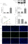
Oe-DNASE1L3 inhibits migration, invasion and tube formation of A549 cells. (A) Migration of A549 cells transfected with Oe-DNASE1L3 was detected by wound healing assay. Magnification, x100. (B) Invasion of A549 cells transfected with Oe-DNASE1L3 was detected by Transwell assay. Magnification, x100. (C) Tube formation of human umbilical vein endothelial cells cultured in medium from A549 cells transfected with Oe-DNASE1L3 was observed by tube formation assay. Magnification, x40. (D) Expression of epithelial-mesenchymal transformation- and tube formation-associated proteins in A549 cells transfected with Oe-DNASE1L3 was detected by western blotting. ***P<0.001 vs. control. ##P<0.01, ###P<0.001 vs. Oe-NC. DNASE1L3, deoxyribonuclease 1-like 3; Oe, overexpression; NC, negative control; VEGF, vascular endothelial growth factor.
Figure 4
Figure 4
Transcription factor FOXP2 positively regulates DNASE1L3 transcription in A549 cells. (A) Binding between FOXP2 and the DNASE1L3 promoter region was predicted using the JASPAR database. Expression of FOXP2 in A549 cells was detected by (B) reverse transcription-quantitative PCR and (C) western blotting. ***P<0.001 vs. 16HBE. Expression of FOXP2 in A549 cells transfected with Oe-FOXP2, si-FOXP2#1 or si-FOXP2#2 was detected by (D) reverse transcription-quantitative PCR and (E) western blotting. ***P<0.001 vs. control. ###P<0.001 vs. Oe-NC. @@@P<0.001 vs. si-NC. (F) Luciferase activity of A549 cells transfected with DNASE1L3-WT and Oe-FOXP2 was detected by dual-luciferase reporter assay. ***P<0.001 vs. DNASE1L3-WT + Oe-NC. (G) Binding of FOXP2 and DNASE1L3 was confirmed by ChIP. ###P<0.001 vs. IgG. Expression of DNASE1L3 in A549 cells transfected with Oe-FOXP2 or si-FOXP2 was detected by (H) reverse transcription-quantitative PCR and (I) western blotting. ***P<0.001 vs. control. ###P<0.001 vs. Oe-NC. @@@P<0.001 vs. si-NC. DNASE1L3, deoxyribonuclease 1-like 3; FOXP2, forkhead-box P2; Oe, overexpression; NC, negative control; si, small interfering; WT, wild-type; MUT, mutant.
Figure 5
Figure 5
Transcription factor FOXP2 regulates transcription of DNASE1L3 and promotes the proliferation of A549 cells. (A) Viability of A549 cells transfected with si-FOXP2 and Oe-DNASE1L3 was determined by MTT. Proliferation of A549 cells transfected with si-FOXP2 and Oe-DNASE1L3 was (B) observed and (C) quantified by EdU staining. Magnification, x200. *P<0.05, **P<0.01 vs. control, ***P<0.001. ###P<0.001 vs. si-NC. @@@P<0.001 vs. si-FOXP2+Oe-NC. DNASE1L3, deoxyribonuclease 1-like 3; FOXP2, forkhead-box P2; OD, optical density; Oe, overexpression; NC, negative control; si, small interfering.
Figure 6
Figure 6
Transcription factor FOXP2 regulates transcription of DNASE1L3 and promotes migration, invasion and tube formation of A549 cells. (A) Migration of A549 cells transfected with si-FOXP2 and Oe-DNASE1L3 was detected by wound healing assay. Magnification, x100. (B) Invasion of A549 cells transfected with si-FOXP2 and Oe-DNASE1L3 was determined by Transwell assay. Magnification, x100. (C) Tube formation of human umbilical vein endothelial cells cultured in the medium from A549 cells transfected with si-FOXP2#2 and Oe-DNASE1L3 was observed by tube formation assay. Magnification, x40. (D) Expression of epithelial-mesenchymal transformation- and tube formation-associated proteins in A549 cells transfected with si-FOXP2#2 and Oe-FOXP2 was detected by western blotting. *P<0.05, **P<0.01, ***P<0.001 vs. control. ###P<0.001 vs. si-NC. @@@P<0.001 vs. si-FOXP2#2+Oe-NC. DNASE1L3, deoxyribonuclease 1-like 3; FOXP2, forkhead-box P2; Oe, overexpression; NC, negative control; si, small interfering; VEGF, vascular endothelial growth factor.

References

    1. Sung H, Ferlay J, Siegel RL, Laversanne M, Soerjomataram I, Jemal A, Bray F. Global cancer statistics 2020: GLOBOCAN estimates of incidence and mortality worldwide for 36 cancers in 185 countries. CA Cancer J Clin. 2021;71:209–249. doi: 10.3322/caac.21660. - DOI - PubMed
    1. Reck M, Rabe KF. Precision diagnosis and treatment for advanced non-small-cell lung cancer. N Engl J Med. 2017;377:849–861. doi: 10.1056/NEJMra1703413. - DOI - PubMed
    1. Qu BL, Cai BN, Yu W, Liu F, Huang YR, Ju ZJ, Wang XS, Ou GM, Feng LC. Radiotherapy effects on brain/bone metastatic adenocarcinoma lung cancer and the importance of EGFR mutation test. Neoplasma. 2016;63:158–162. doi: 10.4149/neo_2016_019. - DOI - PubMed
    1. Wang Y, Yang N, Zhang Y, Li L, Han R, Zhu M, Feng M, Chen H, Lizaso A, Qin T, et al. Effective treatment of lung adenocarcinoma harboring EGFR-activating mutation, T790M, and cis-C797S triple mutations by brigatinib and cetuximab combination therapy. J Thorac Oncol. 2020;15:1369–1375. doi: 10.1016/j.jtho.2020.04.014. - DOI - PubMed
    1. Marinelli D, Mazzotta M, Scalera S, Terrenato I, Sperati F, D'Ambrosio L, Pallocca M, Corleone G, Krasniqi E, Pizzuti L, et al. KEAP1-driven co-mutations in lung adenocarcinoma unresponsive to immunotherapy despite high tumor mutational burden. Ann Oncol. 2020;31:1746–1754. doi: 10.1016/j.annonc.2020.08.2105. - DOI - PubMed