. 2023 Jan 6:13:1082890.
doi: 10.3389/fpls.2022.1082890. eCollection 2022.

Multiscale imaging reveals the presence of autophagic vacuoles in developing maize endosperm

Affiliations

Multiscale imaging reveals the presence of autophagic vacuoles in developing maize endosperm

Elsa Arcalís et al. Front Plant Sci. .

Abstract

Cereal endosperm is solely devoted to the storage of proteins and starch that will be used by the embryo upon germination. The high degree of specialization of this tissue is reflected in its endomembrane system, in which ER derived protein bodies and protein storage vacuoles (PSVs) are of particular interest. In maize seeds, the main storage proteins are zeins, that form transport incompetent aggregates within the ER lumen and finally build protein bodies that bud from the ER. In contrast to the zeins, the maize globulins are not very abundant and the vacuolar storage compartment of maize endosperm is not fully described. Whereas in other cereals, including wheat and barley, the PSV serves as the main protein storage compartment, only small, globulin-containing PSVs have been identified in maize so far. We present here a multi-scale set of data, ranging from live-cell imaging to more sophisticated 3D electron microscopy techniques (SBF-SEM), that has allowed us to investigate in detail the vacuoles in maize endosperm cells, including a novel, autophagic vacuole that is present in early developmental stages.

Keywords: autophagy; lytic vacuole; maize endosperm; multiscale imaging; storage vacuole.


Conflict of interest statement

The authors declare that the research was conducted in the absence of any commercial or financial relationships that could be construed as a potential conflict of interest.

Figures
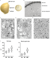
Figure 1
Figure 1
Mapping of the layers in the endosperm. (A) Thin slices of endosperm tissue were obtained immediately above the embryo (arrow). The inset corresponds to the area imaged in B. (B) SEM. Overview of the different endosperm layers. See the aleurone, sub-aleurone and starchy endosperm. A representative cell from the areas marked with the insets 1, 2 and 3 is depicted in C, D and E respectively. (C-E). SEM. Sub-aleurone cell (C), starchy endosperm cell (D), deep-starchy endosperm cell (E). Cell wall (cw), nucleus (n), starch (s), protein storage vacuole (*), zein bodies (arrowheads). (F) Cell and starch size grain in the different endosperm layers. Bars 100 µm (B), 5 µm (C, D), 10 µm (E).
Figure 2
Figure 2
Vacuoles in developing maize seeds. SEM. Developmental stage 2, overview starchy endosperm. Cells contain 4-5 turgent vacuoles (v), that include electrondense globulin inclusions (*). Note the presence of zein bodies in some of them (double arrows). See a single protein storage vacuole (white arrow). Bar 5 µm.
Figure 3
Figure 3
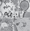
Zein bodies in endosperm cell vacuoles. Developmental stage 2. Starchy endosperm. (A) TEM. Vacuole (v) containing a zein body (arrowhead), globulin inclusions (*) and also some membrane structures (double arrow). Tonoplast (arrow). (B) CLSM, WT seed stained with ER Tracker™ green and neutral red. Several zein bodies (arrowheads) together with globulin inclusions (double arrowheads) within a vacuole (v). White arrows mark zein bodies within the cytoplasma. (C) SBF-SEM. 3D rendering of vacuoles. Zein bodies (green) and globulin inclusions (yellow) within a vacuole. Bars 1 µm (C), 10 µm (B), 1 µm (C).
Figure 4
Figure 4
Vacuoles in different endosperm layers. Developmental stage 2. TEM. (A) Subaleurone, (B) Starchy endosperm, (C) Deep starchy endosperm, (D) Very deep starchy endosperm (see legend in Figure 1 ). Note the presence of zein bodies in the vacuoles (arrowheads) in all layers of the endosperm. Cell wall (cw), mitochondria (m), rER (rough endoplasmic reticulum), starch (s), zein bodies (zb). Bars 2 µm.
Figure 5
Figure 5
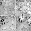
Autophagic vacuoles. Developmental stage 2. Starchy endosperm. (A) CLSM. Neutral red (magenta), MDC (cyan), merged. Vacuoles containing autophagosomes (arrows), autophagosomes within the cytoplasma (arrowheads). (B-F). TEM. Examples of ERphagy (B, C). See the ER fragments (white arrowhead) included in autophagic bodies within the vacuole (v). Examples of mitophagy (D-F). (E) and (F) correspond to the enlargements of insets 1 and 2 respectively found in (D). (E-F). Autophagosomes (arrow) enclosing a mitochondrion (m). (G) SBF-SEM. 3D rendering of vacuoles. See an autophagic body containing a mitochondrion within a vacuole (pale yellow, vacuolar membrane; red, mitochondiron). Globulins (*), protein storage vacuole (PSV), tonoplast (black arrow), zein bodies (zb). Bars 10 µm (A), 1 µm (B-G).
Figure 6
Figure 6
Autophagic pathway into the vacuole. Developmental stage 2, starchy endosperm. TEM. (A) Vacuole (v) containing globulins (*), membrane structures (double white arrowheads) and zein bodies. Note a just incorporated zein body, still preserving a membrane (double black arrowheads) and a zein body without any membrane (arrowhead) in the vacuolar lumen. The inset is enlarged in (B). (B) A phagophore surrounding a zein body (arrowheads). (C) Enlargement of the inset in (B) 1. Edge of the zein body, 2. Protein body membrane, ER derived, see a ribosome (white arrow), 3, 4. Phagophore membranes. (D) Autophagosome including a zein body and an electrondense globulin deposit (arrow). (E) Zein body within a vacuole, still surrounded by an ER membrane (white arrowhead) and an autophagosomal single membrane (double arrowheads). Starch (s), vacuole (v), zein body (zb). Bars 0,5 µm.

Similar articles

Cited by

References

    1. Arcalis E., Hormann-Dietrich U., Zeh L., Stoger E. (2020). 3D electron microscopy gives a clue: Maize zein bodies bud from central areas of ER sheets. Front. Plant Sci. 11, 809. doi: 10.3389/fpls.2020.00809 - DOI - PMC - PubMed
    1. Arcalis E., Ibl V., Peters J., Melnik S., Stoger E. (2014). The dynamic behavior of storage organelles in developing cereal seeds and its impact on the production of recombinant proteins. Front. Plant Sci. 5, 439. doi: 10.3389/fpls.2014.00439 - DOI - PMC - PubMed
    1. Arcalis E., Stadlmann J., Marcel S., Drakakaki G., Winter V., Rodriguez J., et al. . (2010). The changing fate of a secretory glycoprotein in developing maize endosperm. Plant Physiol. 153 (2), 693–702. doi: 10.1104/pp.109.152363 - DOI - PMC - PubMed
    1. Baroux C., Grossniklaus U. (2019). Seeds-an evolutionary innovation underlying reproductive success in flowering plants. Curr. Top. Dev. Biol. 131, 605–642. doi: 10.1016/bs.ctdb.2018.11.017 - DOI - PubMed
    1. Becraft P. W. (2012). Endosperm imprinting: a child custody battle? Curr. Biol. 22 (3), R93–R95. doi: 10.1016/j.cub.2011.12.043 - DOI - PubMed

LinkOut - more resources