. 2022 Dec 29:6:100186.
doi: 10.1016/j.jtauto.2022.100186. eCollection 2023.

Selective induction of thymic stromal lymphopoietin expression by novel nitrogen-containing steroid compounds in PAM-212 cells

Affiliations

Selective induction of thymic stromal lymphopoietin expression by novel nitrogen-containing steroid compounds in PAM-212 cells

Yu Wang et al. J Transl Autoimmun. .

Abstract

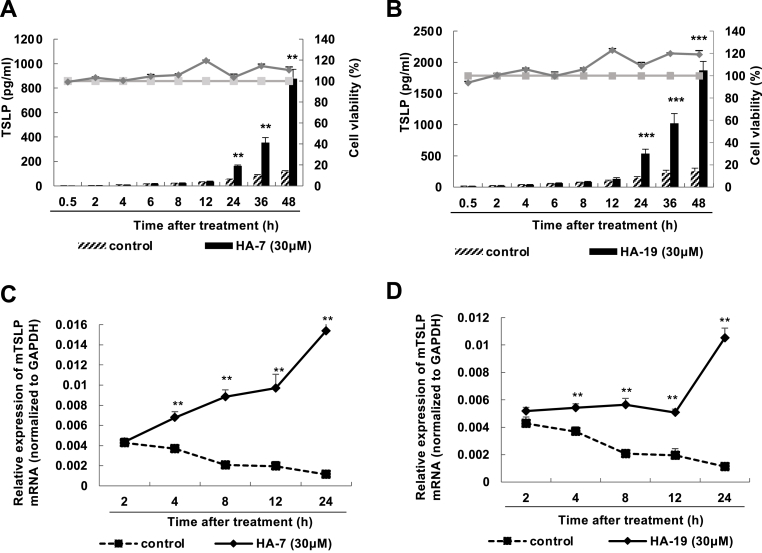
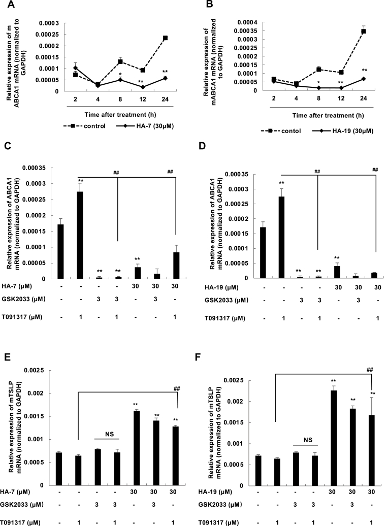
Background: Thymic stromal lymphopoietin (TSLP) has been shown to be able to amplify Tregs. Thus, TSLP induction has the potential to induce endogenous Tregs and control autoimmunity. In the previous research, we found that a new compound named 02F04 can induce TSLP production while simultaneously activating the liver X receptor (LXR). Because LXR activation leads to a decrease in Treg, we attempted to find a 02F04-derivative, druggable lead compound with a basic skeleton that induces TSLP production without activating LXR. As the results, we found HA-7 and HA-19 and, in this study, examined the molecular mechanisms in TSLP production.

Methods: A murine keratinocyte cell line PAM 212 was stimulated with HA-7 and HA-19, and then the expressions of cytokines were examined via ELISA and real-time fluorescence quantitative PCR.

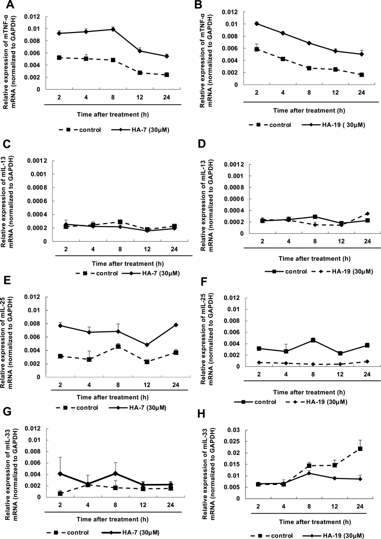
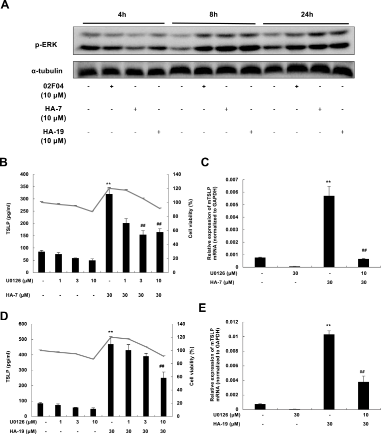
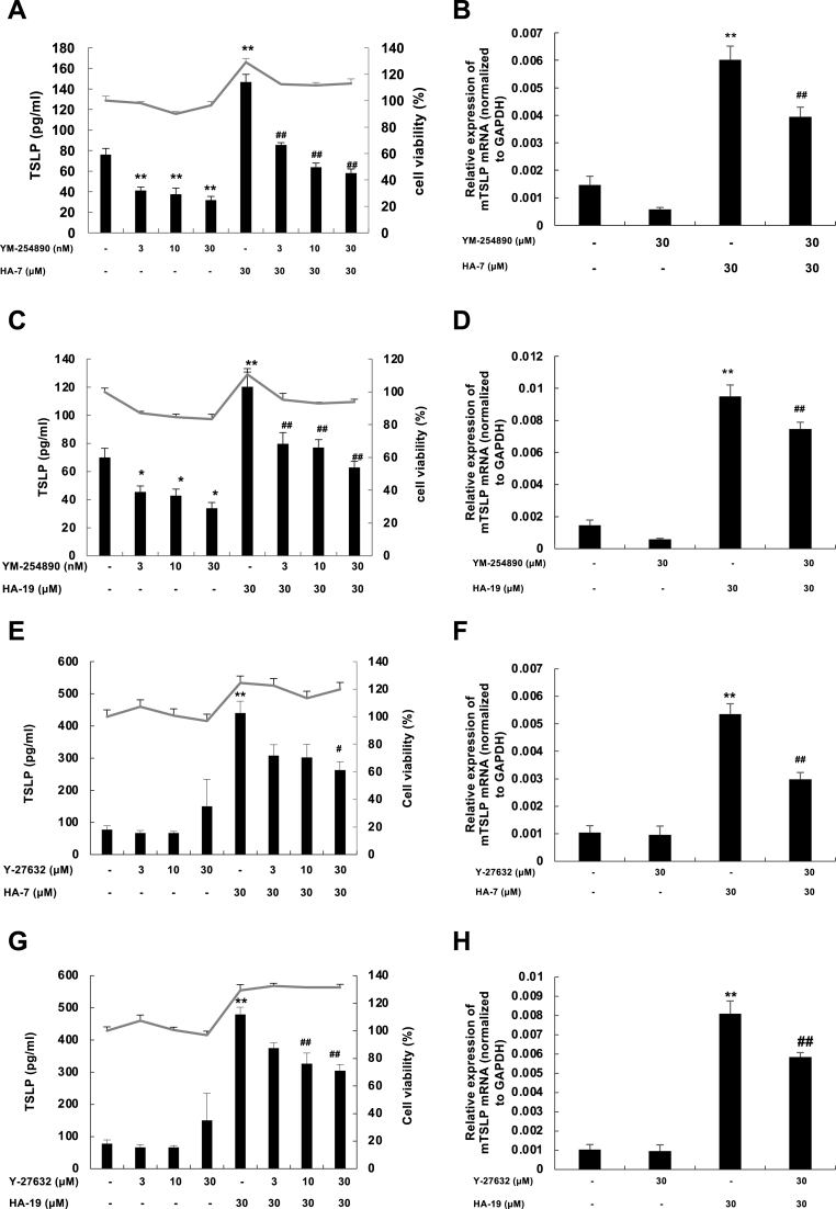
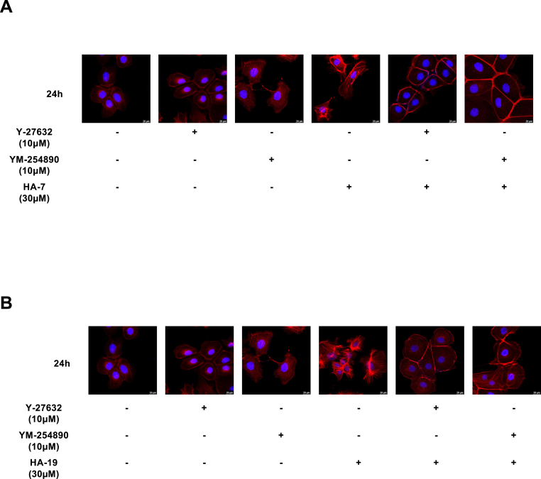
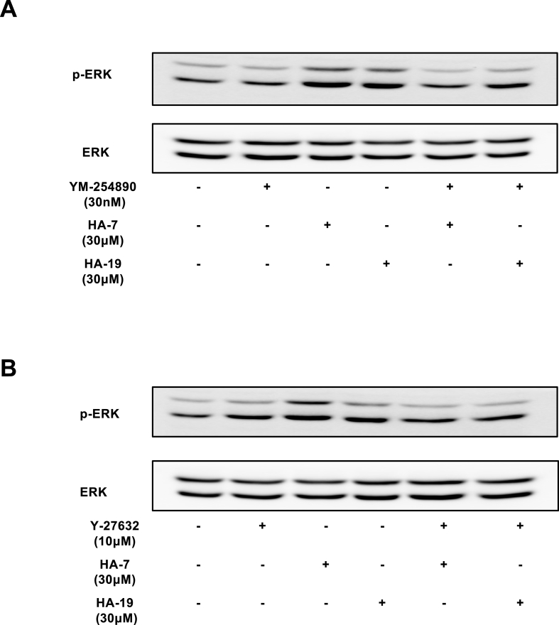
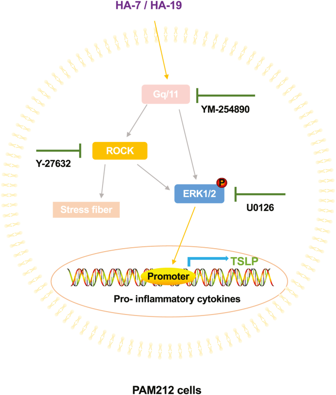
Results: HA-7 and HA-19 induced TSLP production but almost not the expression of TNF-α, IL-13, IL-25, and IL-33 in PAM212 cells. These compounds inhibited LXR activities. The TSLP expression induced by HA-7 and HA-19 was inhibited by the Gq/11 inhibitor YM-254890, ROCK inhibitor Y-27632, and ERK inhibitor U0126. HA-7 and HA-19 also induced the formation of stress fiber and ERK phosphorylation, which were inhibited by YM-254890 and Y-27632.

Conclusions: Our findings indicated that HA-7 and HA-19 selectively induced TSLP production in PAM212 via Gq/11, Rho/ROCK and ERK pathways. Our findings also indicated that TSLP expression was differentially regulated from other cytokines, and the selective expression could be induced with low-molecular-weight compounds such as HA-7 and HA-19.


Figures

Fig. 1
Fig. 1
The chemical structures of 02F04, HA-7 and HA-19 and their inducing effects on TSLP protein in PAM212 cells. The chemical structures of 02F04 (A), HA-7 (B), and HA-19 (C). PAM212 cells were stimulated with 02F04 (D), HA-7 (E), or HA-19 (F) at the indicated concentrations for 24 h. TSLP levels in the culture supernatants were determined by ELISA, and the cytotoxicity test was performed through an MTT assay. Data are presented as means ± SEMs (n = 4). **p < 0.01 and ***p < 0.001 vs. the corresponding control.
Fig. 2
Fig. 2
Time changes in HA-7 and HA-19-induced TSLP production. PAM212 cells were stimulated with HA-7 (A and C) and HA-19 (B and D) at a concentration of 30 μM and at specified times. TSLP levels in the culture supernatants were determined by ELISA, and the cytotoxicity test was performed through an MTT assay (A and B). The mRNA levels of TSLP was determined by qRT-PCR (C and D). Data are presented as means ± SEMs (n = 4). **p < 0.01 and ***p < 0.001 vs. the corresponding control.
Fig. 3
Fig. 3
Effect of HA-7 and HA-19-induced cytokine expression in PAM212 cells. PAM212 cells were stimulated with HA-7 (A, C, E, and G) or HA-19 (B, D, F, and H) at a concentration of 30 μM and at specified times. The mRNA levels of each cytokine were determined by qRT-PCR. Data are presented as means ± SEMs (n = 4).
Fig. 4
Fig. 4
HA-7 and HA-19 inhibit the expression of ABCA1 in LXR. PAM212 cells were stimulated with HA-7 (A) or HA-19 (B) at a concentration of 30 μM and at specified times. The mRNA levels of ABCA1 were determined by qRT-PCR (A and B). The cells were treated with GSK2033 at 3 μM and T091317 at 1 μM with or without HA-7 (C and E) or HA-19 (D and F) at 30 μM for 24 h. The mRNA expressions of ABCA1 (C and D) and TSLP (E and F) were determined by qRT-PCR. Data are presented as means ± SEMs (n = 4). **p < 0.01 vs. the non-stimulated group; ##p < 0.01 vs. T0901317 alone.
Fig. 5
Fig. 5
HA-7 and HA-19 induced ERK phosphorylation and the effect of U0126 on TSLP expression. PAM212 cells were stimulated with 02F04, HA-7 and HA-19 at 10 μM for 4, 8, and 24 h (A). ERK1/2 phosphorylation was measured by western blotting. (B-E) PAM212 cells were stimulated with HA-7 (B and C) or HA-19 (D and E) at 30 μM for 24 h with or without U0126 at the indicated concentrations. TSLP proteins in the culture supernatant and the cytotoxicity (B and D) were determined by ELISA and an MTT assay, respectively. The mRNA levels of TSLP (C and E) were determined by qRT-PCR. Data are presented as means ± SEMs (n = 4). *p < 0.05 and **p < 0.01 vs. the non-stimulated group; ##p < 0.01 vs. HA-7 or HA-19 alone.
Fig. 6
Fig. 6
Effect of YM-254890 and Y-27632 on HA-7- and HA-19-induced TSLP expression. PAM212 cells were stimulated with HA-7 (A, B, E, and F) or HA-19 (C, D, G, and H) at 30 μM for 24 h with or without YM-254890 (A–D) or Y-27632 (E–H) at the indicated concentrations. TSLP proteins in the culture supernatant (A, C, E, and G) were determined by ELISA, and the cytotoxicity test was performed using an MTT assay. The mRNA levels of TSLP (B, D, F, and H) were determined by qRT-PCR. Data are present as means ± SEMs (n = 4). *p < 0.05 and **p < 0.01 vs. the non-stimulated group; ##p < 0.01 vs. HA-7 or HA-19 alone.
Fig. 7
Fig. 7
Effect of YM-254890 and Y-27632 on HA-7- or HA-19-induced polymerization of actin fibers. PAM212 cells were stimulated with HA-7 (A) or HA-19 (B) at 30 μM for 24 h with or without YM-254890 at 10 μM or Y27632 at 10 μM. F-actin and nuclei were stained with rhodamine phalloidin and DAPI, respectively. Immunofluorescence images were obtained using a confocal laser scanning microscope.
Fig. 8
Fig. 8
Effect of YM-254890 and Y-27632 on HA-7- or HA-19-induced ERK phosphorylation. PAM212 cells were stimulated with HA-7 or HA-19 at 30 μM for 24 h with or without YM-254890 at 10 μM (A) or Y27632 at 10 μM (B). ERK1/2 phosphorylation was measured by western blotting.
Fig. 9
Fig. 9
TSLP production is induced by HA7 and HA-19 via the Gq/11-ROCK-ERK pathway.

Similar articles

References

    1. Liu Y.J. Thymic stromal lymphopoietin: master switch for allergic inflammation. J. Exp. Med. Feb. 2006;203(2):269–273. doi: 10.1084/JEM.20051745. - DOI - PMC - PubMed
    1. Varricchi G., et al. Thymic stromal lymphopoietin isoforms, inflammatory disorders, and cancer. Front. Immunol. 2018;9 doi: 10.3389/FIMMU.2018.01595. JUL, Jul. - DOI - PMC - PubMed
    1. Ziegler S.F., Roan F., Bell B.D., Stoklasek T.A., Kitajima M., Han H. Vol. 66. Academic Press Inc.; 2013. The biology of thymic stromal lymphopoietin (TSLP) pp. 129–155. (Adv. Pharmacol.). - DOI - PMC - PubMed
    1. Roan F., Obata-Ninomiya K., Ziegler S.F. Epithelial cell–derived cytokines: more than just signaling the alarm. J. Clin. Invest. 2019;1(4):1441–1451, Apr. doi: 10.1172/JCI124606. 129American Society for Clinical Investigation. - DOI - PMC - PubMed
    1. Liang H.E., Reinhardt R.L., Bando J.K., Sullivan B.M., Ho I.C., Locksley R.M. Divergent expression patterns of IL-4 and IL-13 define unique functions in allergic immunity. Nat. Immunol. 2011;13(1):58–66. doi: 10.1038/ni.2182. Dec. 12011 13. - DOI - PMC - PubMed