Probing fMRI brain connectivity and activity changes during emotion regulation by EEG neurofeedback
- PMID: 36684847
- PMCID: PMC9853008
- DOI: 10.3389/fnhum.2022.988890
Probing fMRI brain connectivity and activity changes during emotion regulation by EEG neurofeedback
Abstract
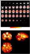
Despite the existence of several emotion regulation studies using neurofeedback, interactions among a small number of regions were evaluated, and therefore, further investigation is needed to understand the interactions of the brain regions involved in emotion regulation. We implemented electroencephalography (EEG) neurofeedback with simultaneous functional magnetic resonance imaging (fMRI) using a modified happiness-inducing task through autobiographical memories to upregulate positive emotion. Then, an explorative analysis of whole brain regions was done to understand the effect of neurofeedback on brain activity and the interaction of whole brain regions involved in emotion regulation. The participants in the control and experimental groups were asked to do emotion regulation while viewing positive images of autobiographical memories and getting sham or real (based on alpha asymmetry) EEG neurofeedback, respectively. The proposed multimodal approach quantified the effects of EEG neurofeedback in changing EEG alpha power, fMRI blood oxygenation level-dependent (BOLD) activity of prefrontal, occipital, parietal, and limbic regions (up to 1.9% increase), and functional connectivity in/between prefrontal, parietal, limbic system, and insula in the experimental group. New connectivity links were identified by comparing the brain functional connectivity between experimental conditions (Upregulation and View blocks) and also by comparing the brain connectivity of the experimental and control groups. Psychometric assessments confirmed significant changes in positive and negative mood states in the experimental group by neurofeedback. Based on the exploratory analysis of activity and connectivity among all brain regions involved in emotion regions, we found significant BOLD and functional connectivity increases due to EEG neurofeedback in the experimental group, but no learning effect was observed in the control group. The results reveal several new connections among brain regions as a result of EEG neurofeedback which can be justified according to emotion regulation models and the role of those regions in emotion regulation and recalling positive autobiographical memories.
Keywords: autobiographical memory; emotion regulation; frontal asymmetry; functional connectivity; neurofeedback; simultaneous recording of EEG and fMRI.
Copyright © 2023 Dehghani, Soltanian-Zadeh and Hossein-Zadeh.
Conflict of interest statement
The authors declare that the research was conducted in the absence of any commercial or financial relationships that could be construed as a potential conflict of interest.
Figures


Similar articles
-
Neural modulation enhancement using connectivity-based EEG neurofeedback with simultaneous fMRI for emotion regulation.Neuroimage. 2023 Oct 1;279:120320. doi: 10.1016/j.neuroimage.2023.120320. Epub 2023 Aug 14. Neuroimage. 2023. PMID: 37586444
-
Global Data-Driven Analysis of Brain Connectivity During Emotion Regulation by Electroencephalography Neurofeedback.Brain Connect. 2020 Aug;10(6):302-315. doi: 10.1089/brain.2019.0734. Epub 2020 Jul 7. Brain Connect. 2020. PMID: 32458692
-
Correlation between amygdala BOLD activity and frontal EEG asymmetry during real-time fMRI neurofeedback training in patients with depression.Neuroimage Clin. 2016 Feb 12;11:224-238. doi: 10.1016/j.nicl.2016.02.003. eCollection 2016. Neuroimage Clin. 2016. PMID: 26958462 Free PMC article.
-
fMRI neurofeedback in emotion regulation: A literature review.Neuroimage. 2019 Jun;193:75-92. doi: 10.1016/j.neuroimage.2019.03.011. Epub 2019 Mar 9. Neuroimage. 2019. PMID: 30862532 Review.
-
Neurofeedback and networks of depression.Dialogues Clin Neurosci. 2014 Mar;16(1):103-12. doi: 10.31887/DCNS.2014.16.1/dlinden. Dialogues Clin Neurosci. 2014. PMID: 24733975 Free PMC article. Review.
Cited by
-
Exploring Neural Mechanisms of Reward Processing Using Coupled Matrix Tensor Factorization: A Simultaneous EEG-fMRI Investigation.Brain Sci. 2023 Mar 13;13(3):485. doi: 10.3390/brainsci13030485. Brain Sci. 2023. PMID: 36979295 Free PMC article.
-
The Use of Transcranial Magnetic Stimulation in Attention Optimization Research: A Review from Basic Theory to Findings in Attention-Deficit/Hyperactivity Disorder and Depression.Life (Basel). 2024 Feb 29;14(3):329. doi: 10.3390/life14030329. Life (Basel). 2024. PMID: 38541654 Free PMC article. Review.
-
Enhancing the accuracy of electroencephalogram-based emotion recognition through Long Short-Term Memory recurrent deep neural networks.Front Hum Neurosci. 2023 Oct 10;17:1174104. doi: 10.3389/fnhum.2023.1174104. eCollection 2023. Front Hum Neurosci. 2023. PMID: 37881690 Free PMC article.
-
Binaural Pulse Modulation (BPM) as an Adjunctive Treatment for Anxiety: A Pilot Study.Brain Sci. 2025 Jan 31;15(2):147. doi: 10.3390/brainsci15020147. Brain Sci. 2025. PMID: 40002480 Free PMC article.
-
Source localization and functional network analysis in emotion cognitive reappraisal with EEG-fMRI integration.Front Hum Neurosci. 2022 Aug 12;16:960784. doi: 10.3389/fnhum.2022.960784. eCollection 2022. Front Hum Neurosci. 2022. PMID: 36034109 Free PMC article.
References
-
- Aday J., Rizer W., Carlson J. M. (2017). “Neural mechanisms of emotions and affect,” in Emotions and affect in human factors and human-computer interaction, ed. Jeon M. (Cambridge, MA: Academic Press; ), 27–87. 10.1016/B978-0-12-801851-4.00002-1 - DOI
LinkOut - more resources
Full Text Sources

