

Case Reports
doi: 10.1016/j.rmcr.2022.101803.
eCollection 2023.
Management of malignancy-induced, life-threatening hypoxemic respiratory failure using a self-expanding Y stent
Affiliations
- PMID: 36685086
- PMCID: PMC9852953
- DOI: 10.1016/j.rmcr.2022.101803
Item in Clipboard
Case Reports
Management of malignancy-induced, life-threatening hypoxemic respiratory failure using a self-expanding Y stent
Respir Med Case Rep.
.
Abstract
We present the case of a young woman transferred to our center with acute hypoxic respiratory failure due to an obstructing subcarinal mass. We review the management and rationale of this respiratory failure at different stages of her hospital course. We describe the approach and rationale in both the intensive care unit as well as the bronchoscopy suite. Finally, we discuss how the use of a novel hybrid Y stent effectively palliated her symptoms.
Keywords: Extracorporeal membrane oxygenation; Hybrid Y stent; Malignant central airway obstruction.
© 2023 The Authors.
Figures
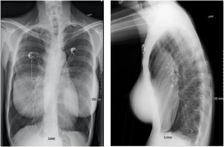
Chest radiograph obtained 1 week prior to emergency transfer. A. PA chest radiograph. Note the lack of clear signs of airway obstruction. B. Lateral chest radiograph. Note the anterior mediastinal fullness and the narrowing of the tracheal airway.
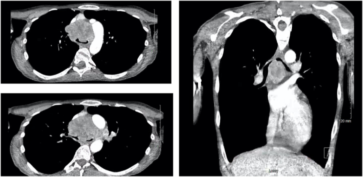
CT imaging prior to transfer and 1 week after the chest radiograph. A. (Top Left) Axial cut at level of aortic arch. B. (Bottom Left) Axial cut just below carina. C. (Right) Representative coronal image.
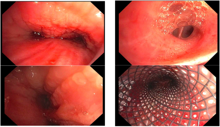
Bronchoscopic images. A. (Top left)Main carina. Tumor infiltration is noted. B. (Bottom left) Left mainstem bronchus at mid bronchus. C. (Top Right) Superior segment bronchus with suspected edema fluid. D. (Bottom Right) Stent within trachea.
References
-
- McGarvey J.M., Pollack C.V. Heliox in airway management. Emerg. Med. Clin. 2008;26(4):905–920. (viii) - PubMed
-
- Ivanick N.M., Moh M., Seeley E.J., Benn B.S. Bilateral endobronchial masses and severe hypoxemic respiratory failure. J. Bronchology Interv. Pulmonol. 2019;26(4):e65–e67. - PubMed
-
- Sakr L., Dutau H. Massive hemoptysis: an update on the role of bronchoscopy in diagnosis and management. Respiration. 2010;80(1):38–58. - PubMed
Publication types
LinkOut - more resources
Full Text Sources
