Review
. 2023;30(3):1495-1552.
doi: 10.1007/s11831-022-09836-2. Epub 2023 Jan 18.

A Review of Image-Based Simulation Applications in High-Value Manufacturing

Affiliations
Review

A Review of Image-Based Simulation Applications in High-Value Manufacturing

Llion Marc Evans et al. Arch Comput Methods Eng. 2023.

Abstract

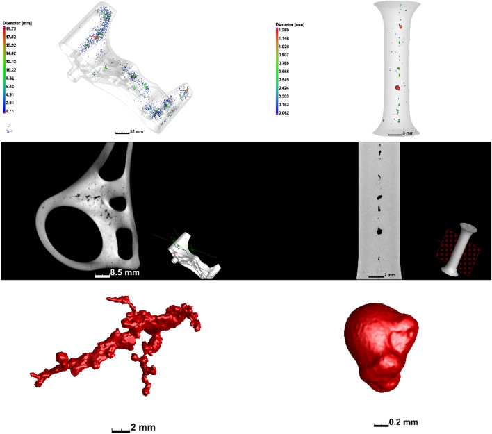
Image-Based Simulation (IBSim) is the process by which a digital representation of a real geometry is generated from image data for the purpose of performing a simulation with greater accuracy than with idealised Computer Aided Design (CAD) based simulations. Whilst IBSim originates in the biomedical field, the wider adoption of imaging for non-destructive testing and evaluation (NDT/NDE) within the High-Value Manufacturing (HVM) sector has allowed wider use of IBSim in recent years. IBSim is invaluable in scenarios where there exists a non-negligible variation between the 'as designed' and 'as manufactured' state of parts. It has also been used for characterisation of geometries too complex to accurately draw with CAD. IBSim simulations are unique to the geometry being imaged, therefore it is possible to perform part-specific virtual testing within batches of manufactured parts. This novel review presents the applications of IBSim within HVM, whereby HVM is the value provided by a manufactured part (or conversely the potential cost should the part fail) rather than the actual cost of manufacturing the part itself. Examples include fibre and aggregate composite materials, additive manufacturing, foams, and interface bonding such as welding. This review is divided into the following sections: Material Characterisation; Characterisation of Manufacturing Techniques; Impact of Deviations from Idealised Design Geometry on Product Design and Performance; Customisation and Personalisation of Products; IBSim in Biomimicry. Finally, conclusions are drawn, and observations made on future trends based on the current state of the literature.


Figures

Fig. 1
Fig. 1
Schematic showing the broad stages for an IBSim workflow which, in this instance, converts X-ray Computed Tomography data into an FEA simulation. This example is a metallic component from a heat exchanger, where the geometry and quality of bonding at the interfaces are integral to the part’s thermal performance
Fig. 2
Fig. 2
Visualisations of a carbon fibre-carbon matrix composite: a photograph, b X-ray radiograph, c volume rendering of XCT data, d 3 orthoslices in the xy, xz and yz planes from XCT data
Fig. 3
Fig. 3
Schematic demonstrating various levels of detail possible when segmenting a complex object. a Photograph of a lemon cut in half and bd image segmented into increasing number of phases: b background, fruit; c background, peel, interior; d background, peel, flesh, seed
Fig. 4
Fig. 4
Flowchart demonstrating the relationship between ‘as designed’ simulations and physical testing conventionally used with more novel ‘as manufactured’ virtual testing with IBSim
Fig. 5
Fig. 5
a A methodology for the development of micromechanical model of asphalt mixtures and simulations (redrawn from [20]); b 2D image of asphalt mixture before FE-meshing [20]; c FEA simulation of image-based heterogenous asphalt mixture under shear load [20]
Fig. 6
Fig. 6
a A schematic for generating virtual specimens based on statistical data of real samples (redrawn from [29]) and b μCT image of a C/SiC woven composite and the centres-of-mass of tow sections [30]; c a 3D rendered virtual specimen [29]
Fig. 7
Fig. 7
a Volumetric rendering from CT data of fibre composites and b CAD rendition of their fibres [33]
Fig. 8
Fig. 8
a 3D CT reconstruction of Ti foam with a unit cell and b its homogenised FEA model under compression applied to the unit cell [48]
Fig. 9
Fig. 9
A schematic of CT-based experimental and numerical investigation of closed-cell metallic foams [49]
Fig. 10
Fig. 10
a CT-based FEA model of nonwoven solid under compression [55] and b a statistical realisation (FEA) model of fibre-glass pack [59]
Fig. 11
Fig. 11
a Pore-space image of Mount Gambier; b its pore-network model extracted/computed from CT images [60]
Fig. 12
Fig. 12
a AM A356/316L composite in low resolution and its unit cell in high resolution with microporosities (316L in dark and A356 in bright contrast) and b the unit cell with interfacial porosity in high resolution [65]
Fig. 13
Fig. 13
a Effect of RVE size on flow permeability for three different porosity levels (E) [75]; b SEM image of nanofibre network and its 3D model [78]
Fig. 14
Fig. 14
a initial surface; b surface after Boolean subtraction operation; d re-meshed surface with triangular elements; e polyhedral volume mesh of porous structure of electrodes; c, f heat transfer analyses over different domain volume size of LiFePO4 electrodes [86]
Fig. 15
Fig. 15
Virtual manufacturing workflow from graphite foam interlayer to CAD pipe and CAD armour with thermal boundary conditions [94]
Fig. 16
Fig. 16
a A computational model of anode microstructure with boundary conditions [104]; b 3D current stream line distribution (red and blue colours are ionic and electronic currents, respectively) [104]; c individual complex particles extracted from reconstructed volume of commercial lithium-ion batteries for stress analysis [105]
Fig. 17
Fig. 17
Examples of casting porosity including (left) shrinkage porosity and (right) gas porosity, image from [120]. The samples are a commercial sand-cast aluminium alloy automotive part (left) and investment cast titanium alloy machined to a tensile dogbone geometry [145]
Fig. 18
Fig. 18
Examples of porosity in metal AM including (left) lack of fusion porosity and (right) keyhole porosity, image from [120]. The samples are small cubes of titanium alloy, manufactured using different process parameters—such cubes are often used to optimise the process allowing up to 99.99% dense parts under optimal conditions []
Fig. 19
Fig. 19
IBSim model before (left) and after (right) tensile testing to failure [145]
Fig. 20
Fig. 20
IBSim model of loading applied to a gyroid lattice structure of titanium alloy produced by L-PBF. The small section viewed here is cropped from the larger structure showing the location of high stresses, and the rough surface exacerbates this [163]. Compressive loading is applied in the vertical direction in the image
Fig. 21
Fig. 21
Reproduced from [174] where heterogeneous solids are grouped and further subdivided by microstructure classes
Fig. 22
Fig. 22
Showing the specific role of each microstructural phase (reproduced from [110])
Fig. 23
Fig. 23
CAD variance analysis of an actual AM bracket (with colour coding showing deviation) compared to its CAD design (shown in yellow mesh). This example is from round robin tests [196] whereby AM parts were analysed by fixed XCT workflows, one of which is to evaluate differences between actual geometry and design geometry
Fig. 24
Fig. 24
Customer involvement and modularity in the production cycle of mass customisation [239]
Fig. 25
Fig. 25
Von-Mises stress distribution of implant, custom abutment, conventional abutment and screw under loading along the abutment long axis (row 1) and along the implant long axis (row 2) [265]
Fig. 26
Fig. 26
Isodose lines calculated by means of IBSim CT simulations of a phantom head a with no bolus, b with a commercial bolus and c a bespoke AM bolus [285]
Fig. 27
Fig. 27
A digital twin reconstructed by the fusion of facial scan and CBCT images. a Coronal view of the face. b Sagittal view of the face [288]
Fig. 28
Fig. 28
FEA analysis showing the stress and temperature analysis of the lower and upper part of the orthosis (top and middle row respectively), and the application of Voronoi patterns to reduce the material in the resulting 3D printed custom orthosis (bottom row) [295]
Fig. 29
Fig. 29
A Process of taking pictures with the bioscanner. B Images obtained with the bioscanner. C Measurement of the exposed intestinal surface dimensions for device design. D Verification of the suitability of the prosthesis by extrusion of the fistulous surface. E Placement of the device on the image of the bioscanned wound to determine the correct adaptation to the patient. F 3D printing of the bioprosthesis [244]
Fig. 30
Fig. 30
a Overview of the Cyber Design and Additive Manufacturing (CDAM) system for custom Ankle Foot Orthoses (AFO). b Illustration of the interaction between the hardware and software systems with the cloud storage system [297]
Fig. 31
Fig. 31
Top: An overview of the schematic representation of the digital workflow with Postoperative CBCT 3D volumetric reconstructions. Bottom: A 3D computer-aided design and planning B FDM printed carbon-reinforced PLA face mask C A professional soccer player with a customized face mask during his sport’s practice session [298]
Fig. 32
Fig. 32
Impact locations of the customized helmet: side, front and top (left) and deviation analysis of a participant’s customised helmet (right) [302]
Fig. 33
Fig. 33
Overview of data collected for the M50 seated FEA male. Left—conventional MRI thigh cross section and lateral view of neck; middle—quasi-seated CT scan and external body laser scan in seated posture; right—full human body model (bone and muscle) [308]
Fig. 34
Fig. 34
a SEM image of chiton’s armour and b segmented X-ray volume; c sketches of parametric CAD model; d comparison between the CT data-based CAD design and the segmented 3D volume of the actual chiton armour components; e virtually buckled FEA model of man-made armour; f a prototype of chiton-inspired armour (reproduced from [313])
Fig. 35
Fig. 35
Design steps to generate a 3D porous scaffold: a selecting the implantable volume zone (or volume of interest), b 3D homogeneous model geometry, c inserting porous trabecular bone like scaffold [335]; d FEA model of a mandible, scaffold and tissue-engineered bone graft implants [222]
Fig. 36
Fig. 36
Steps from the design to manufacturing: a initial and b optimised FEA meshes, c stack of TIFF images, d STL images of the scaffold, and e manufactured scaffolds (redrawn from [219])
Fig. 37
Fig. 37
Flow behaviour over porous scaffold geometry directly obtained from CT images can be achieved with a list of steps: a stack of μCT images, b binarisation and smoothing of the selected volume of interest, c 3D reconstruction and segmentation of the structure, d Boolean operation over the structural geometry to produce the flow domain (i.e., CFD domain), e meshing process of IBSim CFD model, f assigning various boundary conditions (inlet velocity, outlet pressure and wall boundary conditions) [332]
Fig. 38
Fig. 38
Change in publications by year: (area) as a percentage of the total publications considered; (line) absolute number. The area data was found using the search terms (“numerical simulation” OR “computational engineering”); (“tomography”); ‘Both’ denotes, the crossover of these two search terms. The line data was found using the search term ‘(tomography AND “computational engineering”) OR (tomography AND “numerical simulation”) OR (“image-based simulation” OR “image-based modelling”)’. Data collected from Scopus whilst restricting the search to the ‘Engineering’ and ‘Materials Science’ subject areas and the ‘Article’ document type. Data for 2021-2028 are extrapolated using a 3rd order polynomial

References

    1. Withers PJ, Bouman C, Carmignato S, Cnudde V, Grimaldi D, Hagen CK, Maire E, Manley M, Du Plessis A, Stock SR. X-ray computed tomography. Nat Rev Methods Primers. 2021;1:1–21. doi: 10.1038/s43586-021-00015-4. - DOI
    1. Schlüter S, Sheppard A, Brown K, Wildenschild D. Image processing of multiphase images obtained via X-ray microtomography: a review. Water Resour Res. 2014;50:3615–3639. doi: 10.1002/2014WR015256. - DOI
    1. Rizwan I, Haque I, Neubert J. Deep learning approaches to biomedical image segmentation. Inform Med Unlocke. 2020;18:100297. doi: 10.1016/j.imu.2020.100297. - DOI
    1. Livesey F (2006) Confederation of British Industry, University of Cambridge, Institute for Manufacturing, Defining high value manufacturing, CBI, London. http://www.cbi.org.uk/ndbs/positiondoc.nsf/1f08ec61711f29768025672a0055f.... Accessed 7 Apr 2022.
    1. Pieper SD, Laub DRJ, Rosen JM. A finite-element facial model for simulating plastic surgery. Plast Reconstr Surg. 1995;96:1100–1105. doi: 10.1097/00006534-199510000-00014. - DOI - PubMed

LinkOut - more resources