. 2023 Jan 4:13:1055234.
doi: 10.3389/fphys.2022.1055234. eCollection 2022.

Placental treatment with insulin-like growth factor 1 via nanoparticle differentially impacts vascular remodeling factors in guinea pig sub-placenta/decidua

Affiliations

Placental treatment with insulin-like growth factor 1 via nanoparticle differentially impacts vascular remodeling factors in guinea pig sub-placenta/decidua

Baylea N Davenport et al. Front Physiol. .

Abstract

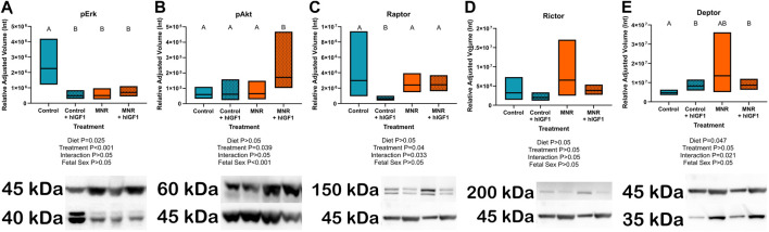
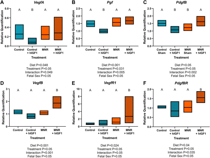
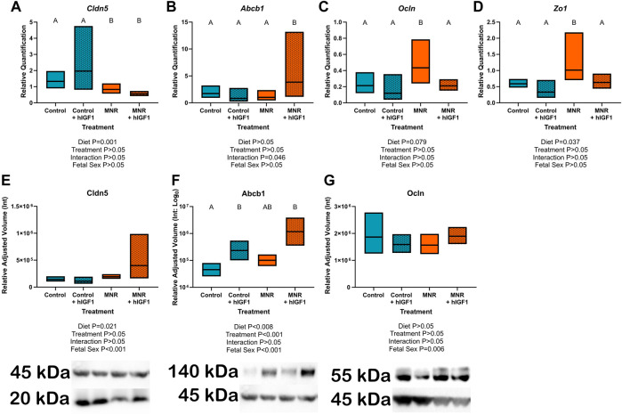
Clinically, fetal growth restriction (FGR) is only detectable in later gestation, despite pathophysiological establishment likely earlier in pregnancy. Additionally, there are no effective in utero treatment options for FGR. We have developed a nanoparticle to deliver human insulin-like 1 growth factor (hIGF-1) in a trophoblast-specific manner which results in increased expression of hIGF-1. IGF-1 signaling in the placenta regulates multiple developmental processes including trophoblast invasion and maternal vascular remodeling, both of which can be diminished in the FGR placenta. We aimed to determine the effects of short-term hIGF-1 nanoparticle treatment on sub-placenta/decidua trophoblast signaling mechanisms in FGR and under normal growth conditions. Using the guinea pig maternal nutrient restriction (MNR) model of FGR, ultrasound-guided, intra-placenta injections of hIGF-1 nanoparticle were performed at gestational day 30-33, and dams sacrificed 5 days later. Sub-placenta/decidua tissue was separated from placenta for further analyses. Western blot was used to analyze protein expression of ERK/AKT/mTOR signaling proteins (phospho-Erk (pERK), phospho-Akt (pAKT), raptor, rictor and deptor). qPCR was used to analyze gene expression of vascular/remodeling factors [vascular endothelial growth factor (Vegf), placenta growth factor (Pgf), platelet-derived growth factor (Pdgf)) and tight junction/adhesion proteins (claudin 5 (Cldn5), p-glycoprotein (Abcb1), occludin (Ocln) and tight junction protein 1 (Zo1)]. MNR reduced expression of pERK, PdgfB and Cldn5, and increased expression of Ocln and Zo1 in the sub-placenta/decidua. In MNR + hIGF1 nanoparticle sub-placenta/decidua, expression of PdgfB, Ocln and Zo1 was normalized, whilst pAkt, VegfB, Vegf receptor 1 and PdgfB receptor were increased compared to MNR. In contrast, hIGF-1 nanoparticle treatment of normal placentas reduced expression of pERK, raptor and increased expression of the mTOR inhibitor deptor. This was associated with reduced expression of VegfA, Plgf, and PdgfB. Here we have shown that the impact of hIGF-1 nanoparticle treatment is dependent on pregnancy environment. Under MNR/FGR, hIGF-1 nanoparticle treatment triggers increased expression of growth factors and normalization of EMT factors. However, under normal conditions, the response of the placenta is to decrease AKT/mTOR signaling and growth factor expression to achieve homeostasis.

Keywords: fetal growth restriction; insulin-like growth factor 1; placenta; pregnancy; therapeutic.


Conflict of interest statement

The authors declare that the research was conducted in the absence of any commercial or financial relationships that could be construed as a potential conflict of interest.

Figures

FIGURE 1
FIGURE 1
Effect of maternal nutrient restriction (MNR) diet and hIGF-1 nanoparticle treatment on expression of Erk/Akt signaling proteins. (A) Protein expression of phospho-Erk (pErk) was reduced in Control + hIGF-1, MNR and MNR + hIGF-1 sub-placenta/decidua when compared to Control. (B) In MNR + hIGF-1 nanoparticle treatment there was increased expression of phospho-Akt (pAkt) compared to MNR, but there was no difference between Control + hIGF-1 and Control sub-placenta/decidua. (C) Compared to Control, there was decreased expression of Raptor in Control + hIGF-1 nanoparticle treatment, but no difference with MNR or MNR + hIGF-1 nanoparticle treatment. (D) There was no difference in the expression of Rictor across any groups. (E) hIGF-1 nanoparticle treatment increased expression of Deptor in both Control + hIGF-1 and MNR + hIGF-1 sub-placenta decidua when compared to Control. Representative western blots for each protein are shown below the graphs with the beta-actin loading control (45 kDa). Western blots for Raptor, Rictor and Deptor were performed on the same membrane, therefore the beta-actin loading control is the same. Samples loaded left to right: Control, Control + hIGF-1, MNR, MNR + hIGF-1. n = 4 Control dams (8 sub-placenta/decidua), 4 Control + hIGF-1 dams (8 sub-placenta/decidua) 4 MNR dams (8 sub-placenta/decidua) and 4 MNR + hIGF-1 dams (8 sub-placenta/decidua). Data are estimated marginal means ±95% confidence interval. p values calculated using generalized estimating equations with Bonferroni post hoc analysis. Different letters denote a significant difference of p ≤ .05.
FIGURE 2
FIGURE 2
Effect of maternal nutrient restriction (MNR) diet and hIGF-1 nanoparticle treatment on expression of angiogenic growth factors. Compared to Control, hIGF-1 nanoparticle treatment reduced mRNA expression of Vascular endothelial growth factor A (VegfA; (A) and Placenta growth factor (Pgf; (B) in control + hIGF-1 sub-placenta/decidua but was not different in MNR and MNR + hIGF-1 nanoparticle treatment sub-placenta/decidua. (C). Expression of Platelet-derived growth factor B (PdgfB) was decreased in Control + hIGF-1 and MNR sub-placenta/decidua compared to Control, and increased in MNR + hIGF-1 to normal. In MNR + hIGF-1 nanoparticle treatment, there was increased expression of VegfB (D), Vegf Receptor 1 (VegfR1; (E) and PdgfB Receptor (PdgfBR; (F) compared to all other groups. n = 6 Control dams (12 sub-placenta/decidua), 4 Control + hIGF-1 dams (12 sub-placenta/decidua) 5 MNR dams (10 sub-placenta/decidua) and 7 MNR + hIGF-1 dams (13 sub-placenta/decidua). Data are estimated marginal means ±95% confidence interval. p values calculated using generalized estimating equations with Bonferroni post hoc analysis. Different letters denote a significant difference of p ≤ .05.
FIGURE 3
FIGURE 3
Protein localization of epithelial-mesenchymal transition (EMT) factors in the sub-placenta/decidua. (A) Representative image of immunohistochemistry (IHC) staining of Claudin 5 in the sub-placenta/decidua trophoblasts (closed arrow head) that line maternal spiral arteries. (B) Representative image of IHC staining of Abcb1 (P-glycoprotein) in the sub-placenta/decidua cytotrophoblasts (closed arrow head) of the cell column. (C) Representative image of IHC staining of Occludin in the trophoblast progenitor cells in the cell columns (open arrow head). (D) Representative image of double label IHC staining for Cytokeratin (brown; closed arrow head), indicating trophoblast cells, and Vimentin (black: red asterisk), indicating blood vessels, of a maternal spiral artery. (E) Representative image of double label IHC staining for Cytokeratin (brown; closed arrow head), indicating cytotrophoblast cells, and Vimentin (black: red asterisk), indicating fetal blood vessels, of the sub-placenta cell column. Trophoblast progenitor cells in the cell column stained weakly for cytokeratin (open arrow head). Scale bar = 10 μm n = 3–5 control placentas.
FIGURE 4
FIGURE 4
Effect of maternal nutrient restriction (MNR) diet and hIGF-1 nanoparticle treatment on expression of epithelial-mesenchymal transition factors. (A). There was decreased expression of Claudin 5 (Cldn5) in MNR and MNR + hIGF-1 sub-placenta/decidua compared to Control and Control + hIGF-1 nanoparticle treatment (B). There was increased expression of P-glycoprotein (Abcb1) in MNR + hIGF-1 compared to Control, Control + hIGF-1 and MNR. (C) In MNR sub-placenta/decidua, there was increased expression of Occludin (Ocln; (C) and Tight junction protein 1 (Zo1; (D) compared to Control, but expression was decreased in MNR + hIGF-1 nanoparticle treatment to normal. (E) Neither diet or hIGF-1 nanoparticle treatment affected protein expression of Cldn5. (F) Protein expression of Abcb1 was increased in Control + hIGF-1 and MNR + hIGF-1 nanoparticle compared to control. (G) Neither diet or hIGF-1 nanoparticle treatment affected protein expression of Ocln. Representative western blots for each protein are shown below the graphs with the beta-actin loading control (45 kDa). Western blots for Cldn5 and Abcb1 were performed on the same membrane, therefore the beta-actin loading control is the same. Samples loaded, from left-right: Control, Control + hIGF-1, MNR, MNR + hIGF1. qPCR analysis: n = 6 Control dams (12 sub-placenta/decidua), 4 Control + hIGF-1 dams (12 sub-placenta/decidua) 5 MNR dams (10 sub-placenta/decidua) and 7 MNR + hIGF-1 dams (13 sub-placenta/decidua). Western blot: n = 4 Control dams (8 sub-placenta/decidua), 4 Control + hIGF-1 dams (8 sub-placenta/decidua) 4 MNR dams (8 sub-placenta/decidua) and 4 MNR + hIGF-1 dams (8 sub-placenta/decidua). Data are estimated marginal means ±95% confidence interval. p values calculated using generalized estimating equations with Bonferroni post hoc analysis. Different letters denote a significant difference of p ≤ .05.

Similar articles

Cited by

References

    1. Abbas Y., Turco M. Y., Burton G. J., Moffett A. (2020). Investigation of human trophoblast invasion in vitro . Hum. Reprod. Update 26 (4), 501–513. 10.1093/humupd/dmaa017 - DOI - PMC - PubMed
    1. Abd Ellah N., Taylor L., Troja W., Owens K., Ayres N., Pauletti G. (2015). Development of non-viral, trophoblast-specific gene delivery for placental therapy. PLoS One 10 (10), e0140879. 10.1371/journal.pone.0140879 - DOI - PMC - PubMed
    1. Adu-Gyamfi E. A., Czika A., Gorleku P. N., Ullah A., Panhwar Z., Ruan L. L. (2021). The involvement of cell adhesion molecules, tight junctions, and gap junctions in human placentation. Reprod. Sci. 28 (2), 305–320. 10.1007/s43032-020-00364-7 - DOI - PubMed
    1. Ahokas R. A., Anderson G. D., Lipshitz J. (1983). Cardiac output and uteroplacental blood flow in diet-restricted and diet-repleted pregnant rats. Am. J. Obstet. Gynecol. 146 (1), 6–13. 10.1016/0002-9378(83)90918-3 - DOI - PubMed
    1. Bernstein I. M., Horbar J. D., Badger G. J., Ohlsson A., Golan A. (2000). Morbidity and mortality among very-low-birth-weight neonates with intrauterine growth restriction. The Vermont Oxford Network. Am. J. Obstet. Gynecol. 182 (1 Pt 1), 198–206. 10.1016/s0002-9378(00)70513-8 - DOI - PubMed