

Distribution of intraperitoneally administered deuterium-labeled water in aquaporin-4-knockout mouse brain after middle cerebral artery occlusion
- PMID: 36685250
- PMCID: PMC9853453
- DOI: 10.3389/fnins.2022.1071272
Distribution of intraperitoneally administered deuterium-labeled water in aquaporin-4-knockout mouse brain after middle cerebral artery occlusion
Abstract
Introduction: As the movement of water in the brain is known to be involved in neural activity and various brain pathologies, the ability to assess water dynamics in the brain will be important for the understanding of brain function and the diagnosis and treatment of brain diseases. Aquaporin-4 (AQP4) is a membrane channel protein that is highly expressed in brain astrocytes and is important for the movement of water molecules in the brain.
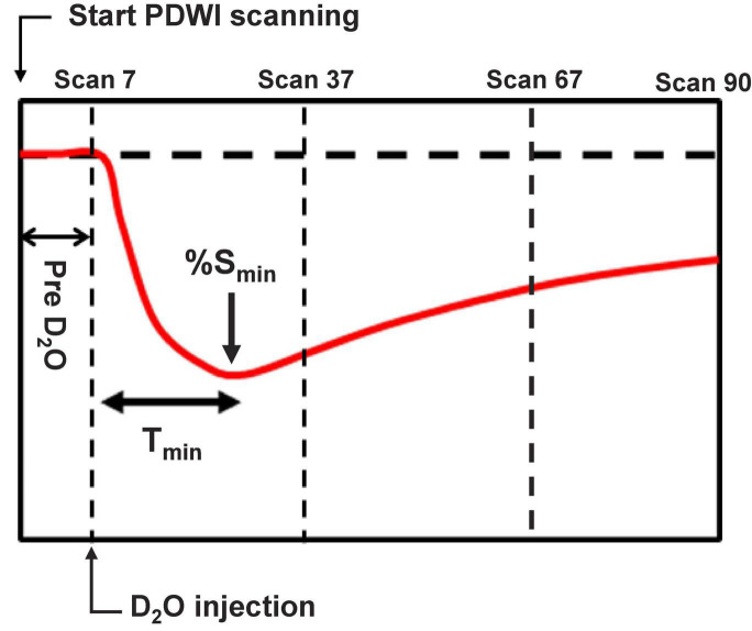
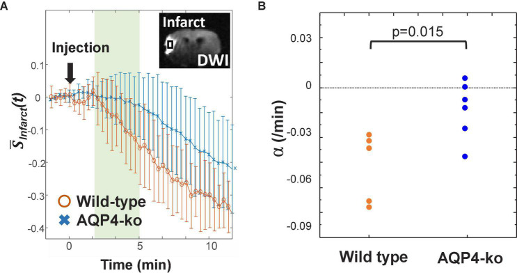
Methods: In this study, we investigated the contribution of AQP4 to brain water dynamics by administering deuterium-labeled water (D2O) intraperitoneally to wild-type and AQP4 knockout (AQP4-ko) mice that had undergone surgical occlusion of the middle cerebral artery (MCA). Water dynamics in the infarct region and on either side of the anterior cerebral artery (ACA) was monitored with proton-density-weighted imaging (PDWI) performed on a 7T animal MRI.
Results: D2O caused a negative signal change quickly after administration. The AQP4-ko mice showed a delay of the time-to-minimum in both the contralateral and ipsilateral ACA regions compared to wild-type mice. Also, only the AQP4- ko mice showed a delay of the time-to-minimum in the ipsilateral ACA region compared to the contralateral side. In only the wild-type mice, the signal minimum in the ipsilateral ACA region was higher than that in the contralateral ACA region. In the infarct region, the signal attenuation was slower for the AQP4-ko mice in comparison to the wild-type mice.
Discussion: These results suggest that AQP4 loss affects water dynamics in the ACA region not only in the infarct region. Dynamic PDWI after D2O administration may be a useful tool for showing the effects of AQP4 in vivo.
Keywords: aquaporin-4; brain ischemia; brain water dynamics; deuterium-labeled water; in vivo proton-density-weighted MRI.
Copyright © 2023 Urushihata, Takuwa, Takahashi, Kershaw, Shibata, Nitta, Tachibana, Yasui, Higuchi and Obata.
Conflict of interest statement
The authors declare that the research was conducted in the absence of any commercial or financial relationships that could be construed as a potential conflict of interest.
Figures


Similar articles
-
Neuroprotective effect of aquaporin-4 deficiency in a mouse model of severe global cerebral ischemia produced by transient 4-vessel occlusion.Neurosci Lett. 2014 Jun 27;574:70-5. doi: 10.1016/j.neulet.2014.03.073. Epub 2014 Apr 6. Neurosci Lett. 2014. PMID: 24717641 Free PMC article.
-
Greatly improved survival and neuroprotection in aquaporin-4-knockout mice following global cerebral ischemia.FASEB J. 2014 Feb;28(2):705-14. doi: 10.1096/fj.13-231274. Epub 2013 Nov 1. FASEB J. 2014. PMID: 24186965 Free PMC article.
-
Reduced brain edema and infarct volume in aquaporin-4 deficient mice after transient focal cerebral ischemia.Neurosci Lett. 2015 Jan 1;584:368-72. doi: 10.1016/j.neulet.2014.10.040. Epub 2014 Nov 1. Neurosci Lett. 2015. PMID: 25449874 Free PMC article.
-
New insights into water transport and edema in the central nervous system from phenotype analysis of aquaporin-4 null mice.Neuroscience. 2004;129(4):983-91. doi: 10.1016/j.neuroscience.2004.06.088. Neuroscience. 2004. PMID: 15561413 Review.
-
Three distinct roles of aquaporin-4 in brain function revealed by knockout mice.Biochim Biophys Acta. 2006 Aug;1758(8):1085-93. doi: 10.1016/j.bbamem.2006.02.018. Epub 2006 Mar 10. Biochim Biophys Acta. 2006. PMID: 16564496 Review.
References
LinkOut - more resources
Full Text Sources
Research Materials