. 2023 Jan 10;9(1):e12892.
doi: 10.1016/j.heliyon.2023.e12892. eCollection 2023 Jan.

Expression, purification, and functional characterization of soluble recombinant full-length simian immunodeficiency virus (SIV) Pr55Gag

Affiliations

Expression, purification, and functional characterization of soluble recombinant full-length simian immunodeficiency virus (SIV) Pr55Gag

Vineeta N Pillai et al. Heliyon. .

Abstract

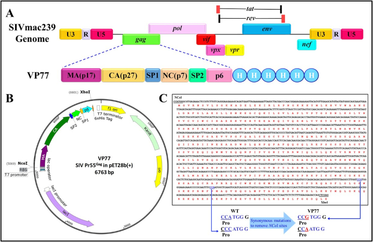
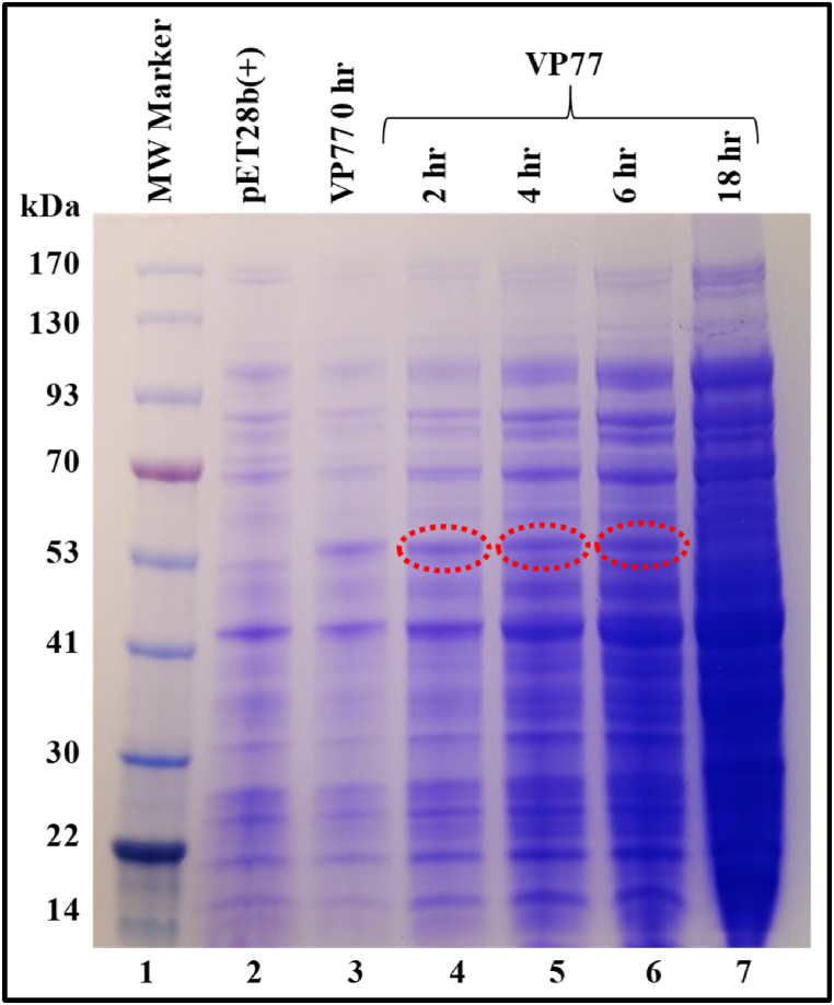
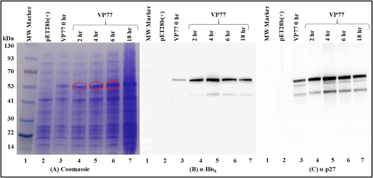
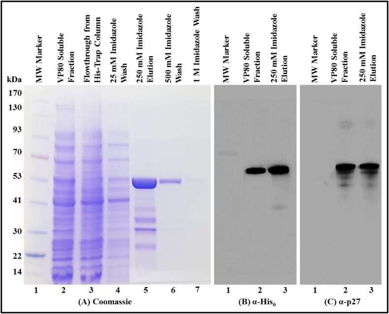
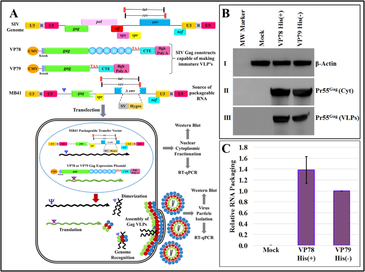
The simian immunodeficiency virus (SIV) precursor polypeptide Pr55Gag drives viral assembly and facilitates specific recognition and packaging of the SIV genomic RNA (gRNA) into viral particles. While several studies have tried to elucidate the role of SIV Pr55Gag by expressing its different components independently, studies using full-length SIV Pr55Gag have not been conducted, primarily due to the unavailability of purified and biologically active full-length SIV Pr55Gag. We successfully expressed soluble, full-length SIV Pr55Gag with His6-tag in bacteria and purified it using affinity and gel filtration chromatography. In the process, we identified within Gag, a second in-frame start codon downstream of a putative Shine-Dalgarno-like sequence resulting in an additional truncated form of Gag. Synonymously mutating this sequence allowed expression of full-length Gag in its native form. The purified Gag assembled into virus-like particles (VLPs) in vitro in the presence of nucleic acids, revealing its biological functionality. In vivo experiments also confirmed formation of functional VLPs, and quantitative reverse transcriptase PCR demonstrated efficient packaging of SIV gRNA by these VLPs. The methodology we employed ensured the availability of >95% pure, biologically active, full-length SIV Pr55Gag which should facilitate future studies to understand protein structure and RNA-protein interactions involved during SIV gRNA packaging.

Keywords: Chromatography; In vitro and in vivo viral particle assembly; Protein purification and expression; RNA binding protein; RNA packaging; Retroviruses; SIV Pr55Gag His6-tagged fusion protein purification; Simian immunodeficiency virus (SIV).


Conflict of interest statement

The authors declare no competing interests.

Figures

Fig. 1
Fig. 1
Construction of VP77 recombinant full-length SIV Pr55Gagbacterial expression vector. (A) Schematic representation of the SIV genome and SIV full-length Pr55Gag bacterial expression plasmid VP77, with the domain organization of Gag and His6-tag. (B) Illustration of VP77, the SIV Pr55Gag expression plasmid constructed using pET28b(+) where Pr55Gag is expressed from the bacteriophage T7 promoter. (C) The translated sequence of SIV Pr55Gag depicts the synonymous mutations (in red) that were introduced in the sequence to inactivate 2 inherent NcoI sites (in blue), for the ease of cloning. (For interpretation of the references to colour in this figure legend, the reader is referred to the Web version of this article.)
Fig. 2
Fig. 2
Expression of the recombinant SIV full-length Pr55Gag-His6-tag fusion protein from E. coli total bacterial cell lysates. Coomassie Brilliant Blue-stained SDS polyacrylamide gel showing expression of recombinant full-length SIV Pr55Gag from total cell lysates at 0, 2, 4, 6 and 18 h after induction with IPTG and cultured at 28 °C. The red dashed circles identify the recombinant protein bands. The uncropped gel image is provided as Supplementary Fig. 1. (For interpretation of the references to colour in this figure legend, the reader is referred to the Web version of this article.)
Fig. 3
Fig. 3
Recombinant SIV full-length Pr55Gag-His6-tag fusion protein expression in the soluble fraction of E. coli. (A) SDS-PAGE gel showing recombinant SIV Pr55Gag-His6-tag fusion protein expression in the soluble fraction at varying time points after suboptimally inducing the bacterial cultures at 28 °C. The red dashed circles identify the recombinant protein bands. Western blot analysis of the soluble fraction confirmed the expression of SIV Pr55Gag-His6-tag fusion protein at varying time points when analyzed with, (B) α-His6 and, (C) with SIV α-p27 monoclonal antibodies respectively, using equal amounts of protein in each lane. The uncropped gel and western blots are provided in Supplementary Fig. 2. (For interpretation of the references to colour in this figure legend, the reader is referred to the Web version of this article.)
Fig. 4
Fig. 4
Synonymous mutations in the Shine-Dalgarno-like sequence to create VP80 recombinant full-length SIV Pr55Gag. (A) VP77 recombinant full-length SIV Pr55Gag showing the Shine-Dalgarno-like sequence (underlined) which is 9 nucleotides upstream of the second in-frame initiation codon ATG (nucleotide 1660, in pink) and, its corresponding amino acids (shown below the sequence). (B) Table listing the predicted translation initiation rates from the actual start codon (in blue) and the second in-frame start codon (in pink) for the wild type VP77 followed by potential synonymous mutations (in red) in the region containing the Shine-Dalgarno-like sequence to disrupt it and inhibit translation from the second in-frame start codon. The mutation which yielded the least translation initiation rate from the second start codon was named VP80. (C) Graphical comparison of the predicted translation rates from the actual start codon (blue) and, the second in-frame start codon (pink), in the wild type VP77 and mutant VP80 containing the altered Shine-Dalgarno-like sequence in recombinant full-length SIV Pr55Gag. (For interpretation of the references to colour in this figure legend, the reader is referred to the Web version of this article.)
Fig. 5
Fig. 5
Immobilized metal affinity chromatography (IMAC) purification of recombinant SIV Pr55Gag-His6-tag fusion protein from the soluble fraction and its Western blot analysis. (A) Coomassie Brilliant Blue-stained SDS-polyacrylamide gel with fractions obtained during IMAC purification of the VP80 SIV Pr55Gag-His6-tag protein: lane 2, soluble fraction; lane 3, HisTrap column flow through; lane 4, after wash with 25 mM imidazole buffer; lane 5, after elution using 250 mM imidazole buffer; lane 6, after wash using 500 mM imidazole buffer; lane 7, after wash with 1 M imidazole buffer. Western blot analysis of the soluble fraction and the elute with 250 Mm imidazole buffer using, (B) α-His6 and, (C) SIV α-p27 monoclonal antibodies, respectively, using equal amounts of protein in each lane. The uncropped gel and western blots are provided in Supplementary Fig. 3. (For interpretation of the references to colour in this figure legend, the reader is referred to the Web version of this article.)
Fig. 6
Fig. 6
Analysis of the fractions obtained by size exclusion chromatography of the recombinant SIV Pr55Gag-His6-tag fusion protein. (A) Chromatogram obtained by plotting the absorbance at 280 nm against the elution time, depicting a distinct peak at between fractions 23 and 29. (B) (a) Coomassie Brilliant Blue-stained SDS-PAGE gel of the fractions 23 to 29 showing resolution of the purified recombinant SIV Pr55Gag expressed from VP80. The cleanest fractions (24–26) were pooled and analyzed by western blotting with (b) α-His6 and, (c) SIV α-p27 monoclonal antibodies respectively, using equal amounts of protein. (C) Absorbance spectra of the final protein preparation on a 220–350 nm range, depicting a 260/280 ratio of 0.6 highlighted in red box. The uncropped gel and western blots are provided in Supplementary Fig. 4. (For interpretation of the references to colour in this figure legend, the reader is referred to the Web version of this article.)
Fig. 7
Fig. 7
Transmission electron micrographs showing VLPs assembled in vitro by bacterially expressed SIV Pr55Gag-His6-tag fusion protein. (AC)In vitro assembled VLPs from grids coated with purified recombinant SIV Pr55Gag-His6-tag fusion protein in the presence of 4% (w/w) yeast tRNA dialyzed overnight at 4 °C at a resolution of 200 nm. (DE) Negative control with in vitro assembly buffer and purified SIV Pr55Gag-His6-tag fusion protein dialyzed in the absence of yeast tRNA overnight at 4 °C, at a resolution of 200 nm. The scale bars represent 200 nm.
Fig. 8
Fig. 8
In vivo assembly of VLPs in eukaryotic cells by SIV Pr55Gag-His6-tag expression plasmid. (AD) Electron micrographs showing SIV Pr55Gag-His6-tag expression plasmid (VP78) transfected into HEK293T cells displaying budding and release of immature VLPs (indicated by red arrows), 72 h post transfection. The electron dense Gag layer is seen accumulating under the plasma membrane and budding out to form VLPs. (E and F) The negative control containing only the SIV transfer vector MB41 and no packaging construct showed no budding or release of VLPs. The scale bars used represents 500 nm for the wide-field pictures and 100 nm for the insets. (For interpretation of the references to colour in this figure legend, the reader is referred to the Web version of this article.)
Fig. 9
Fig. 9
Two-plasmid genetic complementation assay depicting formation of VLPs in eukaryotic cells by SIV Pr55Gagwith and without His6-tag. (A) Schematic representation of the SIV genome, SIV full-length eukaryotic expression constructs VP78 and VP79, expressing Pr55Gag with and without His6-tag and, the SIV transfer vector MB41 which serves as a source for the packageable RNA. Illustration of the in vivo two-plasmid genetic complementation assay where the HEK293T producer cells are co-transfected with the SIV sub-genomic transfer vector (MB41) and, the recombinant SIV Pr55Gag expression plasmids (VP78 or VP79). These cells then produce VLPs containing MB41-specific RNA due to the presence of the packaging signal (Ψ) on the transfer vector. The cytoplasmic fractions and pelleted VLPs were analyzed for Gag expression by western blotting and transfer vector RNA expression by RT-qPCR. (B) Panels I and II are immunoblots of the cytoplasmic fraction from HEK293T producer cells probed with α-β actin monoclonal antibody at 1:25000 dilution and, SIV α-p27 monoclonal antibody used at 1:100 dilution, respectively. Panel III is the immunoblot of the ultracentrifuged VLPs probed with SIV α-p27 monoclonal antibody at 1:100 dilution. (C) Relative RNA packaging efficiency between the VP78 His(+) and VP79 His(−) SIV Pr55Gag RNAs respectively, obtained after RT-qPCR. The relative RNA packaging efficiency was determined by dividing viral RNA packaging values to the cytoplasmic expression normalized to secreted alkaline phosphatase (SEAP) expression for the respective clones. The difference in packaging efficiency between the two clones was statistically not significant. Mock contains only the transfer vector and no packaging construct; hence, it cannot be packaged into viral particles and cannot be picked upon western blots. The uncropped western blots are provided in Supplementary Fig. 5.

Similar articles

Cited by

References

    1. Goff S.P. Intracellular trafficking of retroviral genomes during the early phase of infection: viral exploitation of cellular pathways. J. Gene Med. 2001;3:517–528. doi: 10.1002/1521-2254. (200111)3:6<517::AID-JGM234>3.0.CO;2-E. - DOI - PubMed
    1. Skalka A.M. 2018. Discovering Retroviruses : Beacons in the Biosphere.
    1. Kestler H., Kodama T., Ringler D., Marthas M., Pedersen N., Lackner A., Regier D., Sehgal P., Daniel M., King N., Desrosiers R. Induction of AIDS in rhesus monkeys by molecularly cloned simian immunodeficiency virus. Science. 1990;248:1109–1112. doi: 10.1126/science.2160735. - DOI - PubMed
    1. Lackner A.A., Veazey R.S. Current concepts in AIDS pathogenesis: insights from the SIV/macaque model. Annu. Rev. Med. 2007;58:461–476. doi: 10.1146/annurev.med.58.082405.094316. - DOI - PubMed
    1. Williams K.C., Burdo T.H. HIV and SIV infection: the role of cellular restriction and immune responses in viral replication and pathogenesis. APMIS. 2009;117:400–412. doi: 10.1111/j.1600-0463.2009.02450.x. - DOI - PMC - PubMed

LinkOut - more resources