CD25 as a unique marker on human basophils in stable-mildly symptomatic allergic asthma
- PMID: 36685514
- PMCID: PMC9849741
- DOI: 10.3389/fimmu.2022.1031268
CD25 as a unique marker on human basophils in stable-mildly symptomatic allergic asthma
Abstract
Background: Basophils in acute asthma exacerbation are activated as evidenced by their increased expression levels of activation markers such as CD203c and CD63. However, whether basophils of allergic asthmatics who are in stable phase and have no asthma exacerbations display a specific and distinctive phenotype from those of healthy individuals has yet to be well characterized.
Objective: We aimed to identify the phenotype of basophils from allergic asthmatics in the stable phase and investigate whether such a phenotype is affected by ex vivo allergen stimulation.
Methods: We determined by flow cytometry, the expression of surface proteins such as CD25, CD32, CD63, CD69, CD203c, and CD300a and intracellular anti-apoptotic proteins BCL-2, BCL-xL, and MCL-1. We investigated these markers in blood basophils obtained from well-characterized patients with stable-mildly symptomatic form of allergic asthma with no asthma exacerbation and from healthy individuals. Moreover, we determined ex vivo CD63, CD69, and CD25 on blood basophils from stable-mildly symptomatic allergic asthmatics upon allergen stimulation.
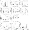
Results: In contrast to all tested markers, CD25 was significantly increased on circulating basophils in the patient cohort with stable-mildly symptomatic allergic asthma than in healthy controls. The expression levels of CD25 on blood basophils showed a tendency to positively correlate with FeNO levels. Notably, CD25 expression was not affected by ex vivo allergen stimulation of blood basophils from stable-mildly symptomatic allergic asthma patients.
Conclusion: Our data identifies CD25 as a unique marker on blood basophils of the stable phase of allergic asthma but not of asthma exacerbation as mimicked by ex vivo allergen stimulation.
Keywords: CD25; basophils; ex vivo stimulation; immunophenotype; stable-mildly symptomatic allergic asthma.
Copyright © 2023 Iype, Rohner, Bachmann, Hermann, Pavlov, von Garnier and Fux.
Conflict of interest statement
The authors declare that the research was conducted in the absence of any commercial or financial relationships that could be construed as a potential conflict of interest.
Figures


Comment on
-
Airway basophils are increased and activated in eosinophilic asthma.Allergy. 2017 Oct;72(10):1532-1539. doi: 10.1111/all.13197. Epub 2017 Jun 14. Allergy. 2017. PMID: 28474352
Similar articles
-
CD203c expression on human basophils is associated with asthma exacerbation.J Allergy Clin Immunol. 2010 Feb;125(2):483-489.e3. doi: 10.1016/j.jaci.2009.10.074. J Allergy Clin Immunol. 2010. PMID: 20159259
-
CD300a is expressed on human basophils and seems to inhibit IgE/FcεRI-dependent anaphylactic degranulation.Cytometry B Clin Cytom. 2012 May;82(3):132-8. doi: 10.1002/cyto.b.21003. Epub 2011 Dec 15. Cytometry B Clin Cytom. 2012. PMID: 22173928
-
Flow cytometry for basophil activation markers: the measurement of CD203c up-regulation is as reliable as CD63 expression in the diagnosis of cat allergy.J Immunol Methods. 2007 Mar 30;320(1-2):40-8. doi: 10.1016/j.jim.2006.12.002. Epub 2007 Jan 3. J Immunol Methods. 2007. PMID: 17275019
-
Allergen-induced basophil activation: CD63 cell expression detected by flow cytometry in patients allergic to Dermatophagoides pteronyssinus and Lolium perenne.Clin Exp Allergy. 2001 Jul;31(7):1007-13. doi: 10.1046/j.1365-2222.2001.01122.x. Clin Exp Allergy. 2001. PMID: 11467990
-
Mast cells and basophils are essential for allergies: mechanisms of allergic inflammation and a proposed procedure for diagnosis.Acta Pharmacol Sin. 2013 Oct;34(10):1270-83. doi: 10.1038/aps.2013.88. Epub 2013 Aug 26. Acta Pharmacol Sin. 2013. PMID: 23974516 Free PMC article. Review.
Cited by
-
CAR-NKT Cells in Asthma: Use of NKT as a Promising Cell for CAR Therapy.Clin Rev Allergy Immunol. 2024 Jun;66(3):328-362. doi: 10.1007/s12016-024-08998-0. Epub 2024 Jul 12. Clin Rev Allergy Immunol. 2024. PMID: 38995478 Review.
-
Advance in the pathogenesis of otitis media with effusion induced by platelet-activating factor.Sci Prog. 2024 Oct-Dec;107(4):368504241265171. doi: 10.1177/00368504241265171. Sci Prog. 2024. PMID: 39380424 Free PMC article. Review.
References
-
- Bischoff SC, de Weck AL, Dahinden CA. Interleukin 3 and granulocyte/macrophage-colony-stimulating factor render human basophils responsive to low concentrations of complement component C3a. Proc Natl Acad Sci United States America (1990) 87(17):6813–7. doi: 10.1073/pnas.87.17.6813 - DOI - PMC - PubMed
Publication types
MeSH terms
Substances
LinkOut - more resources
Full Text Sources
Medical
Research Materials
Miscellaneous

