. 2023 Jan 6:13:1110322.
doi: 10.3389/fimmu.2022.1110322. eCollection 2022.

Cc GSDMEa functions the pore-formation in cytomembrane and the regulation on the secretion of IL-lβ in common carp (Cyprinus carpio haematopterus)

Affiliations

Cc GSDMEa functions the pore-formation in cytomembrane and the regulation on the secretion of IL-lβ in common carp (Cyprinus carpio haematopterus)

Yanjing Zhao et al. Front Immunol. .

Abstract

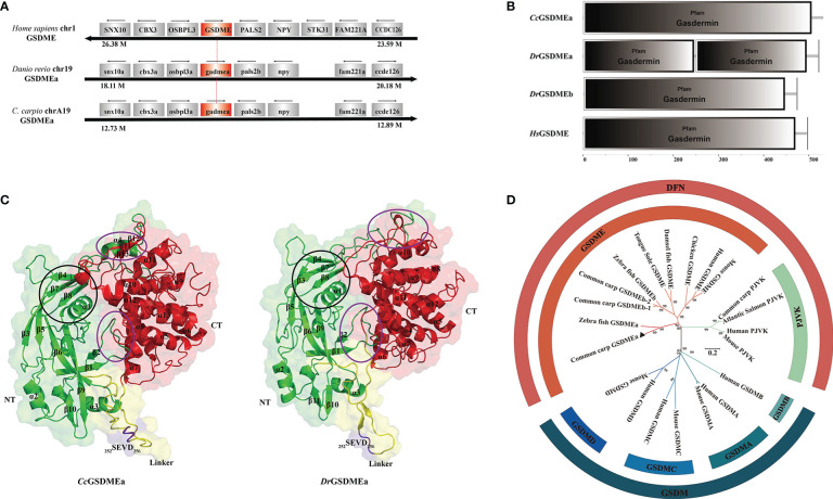
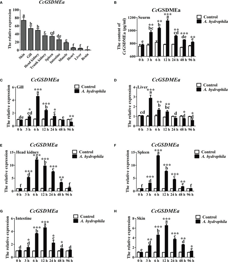
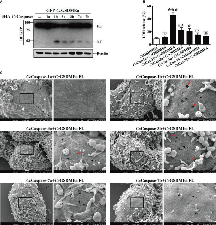
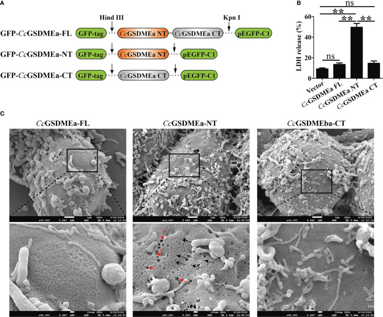
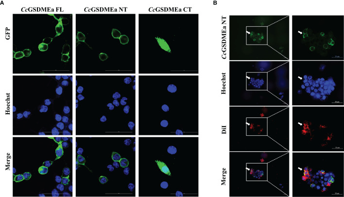
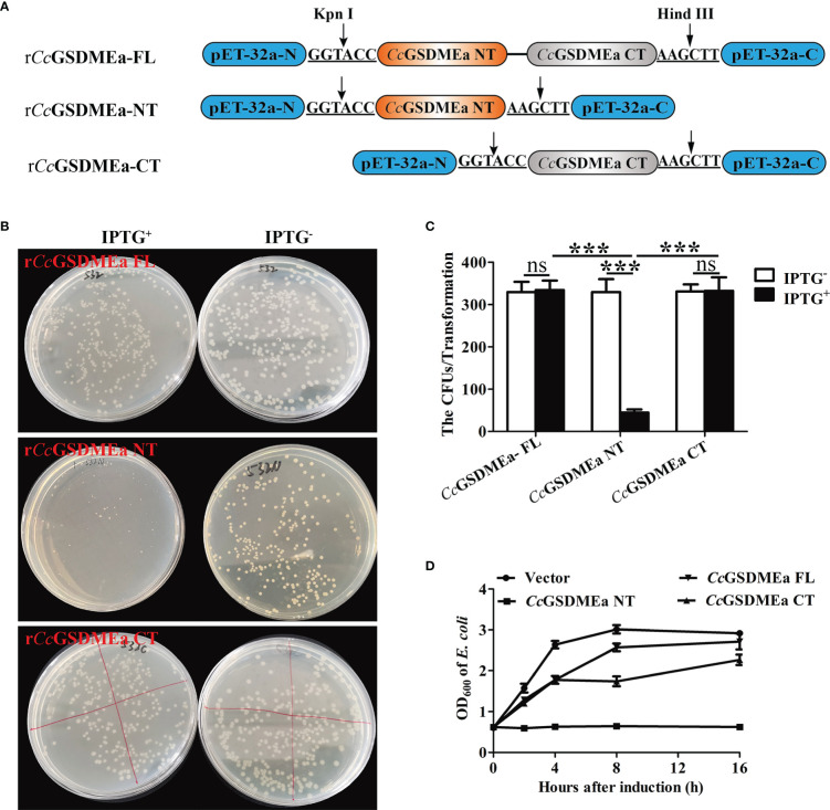
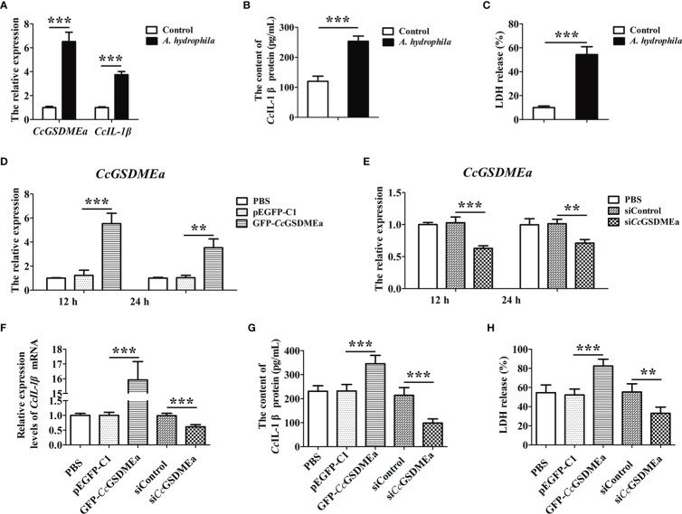
GSDME is the only direct executor of caspase-dependent pyroptosis in both canonical and non-canonical inflammasomes known to date in fish, and plays an important role in anti-bacterial infection and inflammatory response. In order to determine the regulation of GSDMEa on antibacterial infection in innate immune response, the CcGSDMEa gene in common carp (Cyprinus carpio haematopterus) was first identified and characterized, and then its function related to immune defense was investigated. Our results showed that the expressions of CcGSDMEa at the mRNA and protein levels were both significantly increased after Aeromonas hydrophila intraperitoneal infection at the early stage than that in the control group. We found that CcGSDMEa could be cleaved by inflammatory caspase (CcCaspase-1b) and apoptotic caspases (CcCaspase-3a/b and CcCaspase-7a/b). Interestingly, only the CcGSDMEa-NT (1-252 aa) displayed bactericidal activity to Escherichia coli and could punch holes in the membrane of HEK293T cells, whereas CcGSDMEa-FL (1-532 aa) and CcGSDMEa-CT (257-532 aa) showed no above activity and pore-forming ability. Overexpression of CcGSDMEa increased the secretion of CcIL-1β and the release of LDH, and could reduce the A. hydrophila burdens in fish. On the contrary, knockdown of CcGSDMEa reduced the secretion of CcIL-1β and the release of LDH, and could increase the A. hydrophila burdens in fish. Taken together, the elevated expression of CcGSDMEa was a positive immune response to A. hydrophila challenge in fish. CcGSDMEa could perform the pore-formation in cell membrane and the regulation on the secretion of IL-lβ, and further regulate the bacterial clearance in vivo. These results suggested that CcGSDMEa played an important role in immune defense against A. hydrophila and could provide a new insight into understanding the immune mechanism to resist pathogen invasion in teleost.

Keywords: Aeromonas hydrophila; GSDMEa; IL-lβ secretion; caspases; common carp; gasdermin pore.


Conflict of interest statement

The authors declare that the research was conducted in the absence of any commercial or financial relationships that could be construed as a potential conflict of interest.

Figures

Figure 1
Figure 1
Structural feature and phylogenetic evolution of CcGSDMEa. (A) Gene synteny and chromosomal location analysis of CcGSDMEa. The genes adjacent to GSDME loci on human chromosome 1, zebrafish chromosome 19, common carp chromosome A19 are shown. Arrows indicate gene orientation. The small arrows above the gene indicate the direction in which the gene is transcribed. (B) Structure domains of CcGSDMEa, DrGSDMEa/b, and HsGSDME predicted by SMART. (C) The tertiary structures of CcGSDMEa and DrGSDMEa modeled by I-TASSER. The NT domains and CT domains are colored green and red, respectively. The motifs of tetrapeptide 252SEVD256 specifically recognized by caspases are marked in purple, and the linker regions are labeled in yellow. Secondary structure element α-helices and β-strands are indicated in images. The inter-domain interfaces are highlighted by purple ellipses. The α1, β3, β4, β7 and β8 marked by the black circle form the structure of the membrane inserted N-terminal domain. (D) Phylogenetic relationships of GSDM family members. The phylogenetic tree was constructed by MEGA (version 7.0) using the neighbor-joining method. The reliability of each node was estimated by bootstrap test with 1000 replications. The accession numbers of selected GSDM protein sequences from the National Center for Biotechnology Information Reference Sequence database (http://www.ncbi.nlm.nih.gov/RefSeq/) are shown in Table 3 .
Figure 2
Figure 2
Expression profile of CcGSDMEa after A. hydrophila challenge. (A) Tissues distribution of CcGSDMEa in healthy C. carpio. The lowest expression level in brain was chosen as calibration (set as 1), and the relative expression of CcGSDMEa in other tissues was represented as fold-changes to the calibration. (B) The protein expression levels of CcGSDMEa in serum after A. hydrophila challenge. (C-H) The mRNA expression levels of CcGSDMEa in different tissues after A. hydrophila challenge. The control group was chosen as calibration (set as 1). The data were expressed as mean ± SD (n = 3). Significant difference between the different time points were analyzed using one-way ANOVA followed by Tukey’s test, and presented with the different lowercase letters in the group challenged by A. hydrophila (P<0.05), and the same letter was indicated no significant difference (P > 0.05). The significant differences between the A. hydrophila challenge group and the control group at the same time point were analyzed by student’s t test and indicated with asterisks (*, P < 0.05; **, P < 0.01, ***, P < 0.01).
Figure 3
Figure 3
CcGSDMEa-NT was produced from CcGSDMEa cleavage by CcCaspase-1/3/7. (A) The western bolting result that CcGSDMEa was cleaved by CcCaspase-1b/3a/3b/7a/7b. (B) LDH release from the EPC cells co-transfected with CcGSDMEa and the CcCaspase-1a/1b/3a/3b/7a/7b plasmids. (C) Morphological observation of HEK293T cells co-transfected with CcGSDMEa and the CcCaspase-1a/1b/3a/3b/7a/7b plasmids. Red arrows display cell plasma membrane rupture, and dark arrows indicate the gasdermin membrane pores. Values are shown as mean ± SD (n = 3). The LDH release from the cells only transfected with CcGSDMEa was set as control. The significance difference of LDH release between the control cells and the co-transfected cells was analyzed by student’s t test and shown as: ns, P > 0.05; *, P < 0.05; **, P < 0.01; ***, P < 0.001.
Figure 4
Figure 4
Membrane pore formation induced by CcGSDMEa-NT in HEK293T cells. (A) Schematic diagram of the recombinant plasmids of CcGSDMEa-FL (1-532 AA), CcGSDMEa-NT (1-250 AA), CcGSDMEa-CT (257-532 AA) and pEGFP-C1. (B) LDH release from cells transfected with 1000 ng of different forms of CcGSDMEa recombinant plasmids. (C) Morphological observation of HEK293T cells transfected with different forms of CcGSDMEa recombinant plasmids. Red arrows display cell plasma membrane rupture, and dark arrows indicate the membrane pores induced by CcGSDMEa-NT. The LDH release values are shown as M ± SD (n = 3). The significant differences were analyzed by student’s t test, “**” means P < 0.01, “ns” means no significant difference.
Figure 5
Figure 5
Subcellular localizations of CcGSDMEa different forms. (A) Subcellular localizations of CcGSDMEa-FL/NT/CT and Hoechst-stained nucleus in HEK293T cells. (B) Subcellular localization of CcGSDMEa-NT, Hoechst-stained nucleus and DiI-stained cytomembrane in HEK293T cells (white arrowheads). The original microscope images were on the left and the locally enlarged images were on the right in (B).
Figure 6
Figure 6
Bactericidal activity of CcGSDMEa to E. coli. (A) Schematic diagram of the recombinant plasmids of rCcGSDMEa-FL, rCcGSDMEa-NT and rCcGSDMEa-CT. (B) Colony growth of E. coli expressing rCcGSDMEa-FL/NT/CT. (C) Calculation of CFUs on the plates. (D) Growth curves of E. coli expressing rCcGSDMEa-FL/NT/CT after IPTG induction. Values are expressed as M ± SD (n = 3). The statistical difference was analyzed by student’s t test (***, P < 0.001; ns, no significant difference).
Figure 7
Figure 7
CcGSDMEa promotes the expression of CcIL-1β in vivo. (A-D) Overexpression efficiency and knockdown efficiency of CcGSDMEa in gill, liver, spleen, trunk kidney and head kidney of C. carpio. (E, F) The mRNA expression levels of CcIL-1β in the different groups after A. hydrophila challenge. (G, H) The protein expression levels of CcIL-1β in the different groups after A. hydrophila challenge. In each case, the mRNA expression level in the PBS group was chosen as calibration (set as 1). The statistical difference was analyzed by student’s t test (*, P < 0.05; **, P < 0.01; ***, P < 0.001).
Figure 8
Figure 8
CcGSDMEa prevents A. hydrophila colonization in vivo. Bacterial burdens in head kidney (A), spleen (B), gill (C), liver (D) and trunk kidney (E) of different groups infected with A. hydrophila. The data are expressed as mean ± SD (n = 3) and analyzed statistically by student’s t test (*, P < 0.05; **, P < 0.01; ***, P < 0.001).
Figure 9
Figure 9
CcGSDMEa promotes the secretion of CcIL-1β in vitro. (A) The mRNA expression levels of CcGSDMEa and CcIL-1β in EPC cells after A. hydrophila challenge. (B) The content of CcIL-1β protein in the cell culture medium after A. hydrophila challenge. (C) The LDH release in EPC cells after A. hydrophila challenge. (D, E) The overexpression and knockdown efficiency of CcGSDMEa in EPC cells at 12 h and 24 h. (F-H) The mRNA expression levels of CcIL-1β, protein secretion contents of CcIL-1β and the LDH release from cells in overexpression or knockdown group after A. hydrophila challenge. The data were expressed as mean ± SD (n = 3). The significant differences were analyzed using student’s t test (**, P < 0.01; ***, P < 0.001).
Figure 10
Figure 10
The proposed mode of the regulation role of CcGSDMEa on CcIL-1β secretion in common carp.

References

    1. Nakajima T, Hudson MJ, Uchiyama J, Makibayashi K, Zhang J. Common carp aquaculture in neolithic China dates back 8,000 years. Nat Ecol Evol (2019) 3(10):1415–8. doi: 10.1038/s41559-019-0974-3 - DOI - PubMed
    1. Lafferty KD, Harvell CD, Conrad JM, Friedman CS, Kent ML, Kuris AM, et al. Infectious diseases affect marine fisheries and aquaculture economics. Ann Rev Mar Sci (2015) 7:471–96. doi: 10.1146/annurev-marine-010814-015646 - DOI - PubMed
    1. Basu M, Swain B, Maiti NK, Routray P, Samanta M. Inductive expression of toll-like receptor 5 (TLR5) and associated downstream signaling molecules following ligand exposure and bacterial infection in the Indian major carp, mrigal (Cirrhinus mrigala). Fish Shellfish Immunol (2012) 32(1):121–31. doi: 10.1016/j.fsi.2011.10.031 - DOI - PubMed
    1. Orozova P, Sirakov I, Austin DA, Austin B. Recovery of bacillus mycoides, b. pseudomycoides and Aeromonas hydrophila from common carp (Cyprinus carpio) and rainbow trout (Oncorhynchus mykiss) with gill disease. J Fish Dis (2018) 41(1):125–9. doi: 10.1111/jfd.12686 - DOI - PubMed
    1. Di G, Li H, Zhang C, Zhao Y, Zhou C, Naeem S, et al. Label-free proteomic analysis of intestinal mucosa proteins in common carp (Cyprinus carpio) infected with aeromonas hydrophila. Fish Shellfish Immunol (2017) 66:11–25. doi: 10.1016/j.fsi.2017.04.025 - DOI - PubMed

Publication types