. 2022 Dec 22:35:10697.
doi: 10.3389/ti.2022.10697. eCollection 2022.

Formation of Re-Aggregated Neonatal Porcine Islet Clusters Improves In Vitro Function and Transplantation Outcome

Collaborators, Affiliations

Formation of Re-Aggregated Neonatal Porcine Islet Clusters Improves In Vitro Function and Transplantation Outcome

M Honarpisheh et al. Transpl Int. .

Abstract

Neonatal porcine islet-like cell clusters (NPICCs) are a promising source for islet cell transplantation. Excellent islet quality is important to achieve a cure for type 1 diabetes. We investigated formation of cell clusters from dispersed NPICCs on microwell cell culture plates, evaluated the composition of re-aggregated porcine islets (REPIs) and compared in vivo function by transplantation into diabetic NOD-SCID IL2rγ-/- (NSG) mice with native NPICCs. Dissociation of NPICCs into single cells and re-aggregation resulted in the formation of uniform REPI clusters. A higher prevalence of normoglycemia was observed in diabetic NSG mice after transplantation with a limited number (n = 1500) of REPIs (85.7%) versus NPICCs (n = 1500) (33.3%) (p < 0.05). Transplanted REPIs and NPICCs displayed a similar architecture of endocrine and endothelial cells. Intraperitoneal glucose tolerance tests revealed an improved beta cell function after transplantation of 1500 REPIs (AUC glucose 0-120 min 6260 ± 305.3) as compared to transplantation of 3000 native NPICCs (AUC glucose 0-120 min 8073 ± 536.2) (p < 0.01). Re-aggregation of single cells from dissociated NPICCs generates cell clusters with excellent functionality and improved in vivo function as compared to native NPICCs.

Keywords: islet transplantation; neonatal islet-like cell clusters; porcine islets; pseudo-islets; re-aggregated cell clusters; xenotransplantation.


Conflict of interest statement

The authors declare that the research was conducted in the absence of any commercial or financial relationships that could be construed as a potential conflict of interest.

Figures

FIGURE 1
FIGURE 1
Phenotype and architecture of native neonatal porcine islet-like clusters (NPICCs) and re-aggregated porcine islets (REPIs) on day 7 after isolation. (A) Phase contrast pictures of cells cultured in Sphericalplate 5D with 750 cells per microwell (left) and morphology of NPICCs (middle) and REPIs (right) after harvesting. Scale bars, 100 µm. (B) Size distribution of REPIs and NPICCs from n = 5 pancreata. Box plots with median diameter, 25th-75th percentile and minimum and maximum (whiskers). (C) Viability of cells analyzed by FACS (right) and fluorescence microscopy (left) using calcein AM (green, live cells) and propidium iodide (PI) (red, dead cells) dye staining (n = 5). (D) TUNEL assay revealed many TUNEL positive cells (green) in the inner core of large NPICCs. Scale bars, 100 µm (E) Recovery rate of REPIs as percentage of NPICCs (n = 10). (F) Immunofluorescence staining of insulin (red) and glucagon (green) in NPICCs and REPIs (n = 4). Scale bars, 100 µm. (G) Measurement of in vitro beta cell function assessed by glucose stimulated insulin secretion (n = 4). (H) Representative flow cytometric characterization of insulin, glucagon and somatostatin positive cells. (I) Analysis of the percentage of insulin (white bars), glucagon (black bars) and somatostatin (grey bars) positive cells and sum of hormone positive cells (dotted bars) (n = 4). Data are presented as mean ± SD. *p < 0.05, **p < 0.01 vs. NPICCs.
FIGURE 2
FIGURE 2
Transplantation with REPIs improved reversal of diabetes. (A) Kaplan-Meier analysis of time to develop normoglycemia in diabetic NSG mice transplanted with 750 (n = 4) and 1500 REPIs (n = 7) or 750 (n = 4), 1500 (n = 6) and 3000 NPICCs (n = 22) on day 7 after isolation. After transplantation with 1500 REPIs significantly more animals developed normoglycemia defined by non-fasting blood glucose <120 mg/dl compared to the group transplanted with 1500 NPICCs (*p < 0.05). (B–D) Intraperitoneal glucose tolerance test (IPGTT) in diabetic NSG mice transplanted with 1500 REPIs (n = 6) or 3000 NPICCs (n = 10). (B) Glucose response curve during the IPGTT. (C) Glucose clearance during IPGTT assessed by calculating area under the curve (AUC) for glucose 0–120 min was improved in animals transplanted with REPIs (n = 6) **p < 0.01 vs. NPICC group. (D) Insulin secretion at 0 and 10 min after glucose challenge was similar in both transplantation groups. Data are presented as the mean ± SD.
FIGURE 3
FIGURE 3
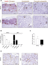
Architecture and revascularization of transplanted grafts. (A) Representative immunohistochemical staining for insulin (red)/glucagon (brown), somatostatin (brown, arrows) and pancreatic polypeptide (PP) (brown, arrrows) in sections of grafted NPICCs and REPIs (1500 IEQ). (B) Quantification of endocrine cells within the grafts revealed a similar proportion of insulin, glucagon and somatostatin cells and a significant lower frequency of PP cells in grafted REPIs (n = 3 per group). **p < 0.01. (C) Quantification of CD31 positive endothelial cells (EC) by immunohistochemistry (n = 3 per group). The results are expressed as CD31 positive area normalized to islet area. (D) Representative images of CD31 staining in grafts. Characteristic CD31 staining (brown) of blood vessels in the grafts. Data are represented as the mean ± SD. Scale bars, 100 µm.

References

    1. Gamble A, Pepper AR, Bruni A, Shapiro AMJ. The Journey of Islet Cell Transplantation and Future Development. Islets (2018) 10(2):80–94. 10.1080/19382014.2018.1428511 - DOI - PMC - PubMed
    1. Giuliani M, Moritz W, Bodmer E, Dindo D, Kugelmeier P, Lehmann R, et al. Central Necrosis in Isolated Hypoxic Human Pancreatic Islets: Evidence for Postisolation Ischemia. Cel Transpl (2005) 14(1):67–76. 10.3727/000000005783983287 - DOI - PubMed
    1. Komatsu H, Cook C, Wang CH, Medrano L, Lin H, Kandeel F, et al. Oxygen Environment and Islet Size Are the Primary Limiting Factors of Isolated Pancreatic Islet Survival. PloS one (2017) 12(8):e0183780. 10.1371/journal.pone.0183780 - DOI - PMC - PubMed
    1. Lehmann R, Zuellig RA, Kugelmeier P, Baenninger PB, Moritz W, Perren A, et al. Superiority of Small Islets in Human Islet Transplantation. Diabetes (2007) 56(3):594–603. 10.2337/db06-0779 - DOI - PubMed
    1. MacGregor RR, Williams SJ, Tong PY, Kover K, Moore WV, Stehno-Bittel L. Small Rat Islets Are superior to Large Islets in In Vitro Function and in Transplantation Outcomes. Am J Physiol Endocrinol Metab (2006) 290(5):E771–9. 10.1152/ajpendo.00097.2005 - DOI - PubMed